

Research Article :
Background: Intraoral digital scanning could offer a win-win
clinical pathway for both doctor and patient, but many factors can affect its
accuracy. This study was designed to investigate the effect of the scanning
number on the precision of digital impression in different gypsum models. With
the development of science and technology, great changes have taken place in
dental medical equipment and technology [1]. Digital technology has been
rapidly disseminated in the field of stomatology [2,3]. Fast and accurate digital impression
acquisition technology is the inevitable trend of future development. Digital
impressions produced by intraoral and extraoral are two entry points for data
acquisition. The extraoral method transfers clinical information to technicians
by scanning impressions or plaster models for prosthetic
reconstruction [4]. But, in the
process of making impression and pouring gypsum model, it is unavoidable to
produce the shrinkage of impression material, expansion in the process of gypsum
hardening and various human
factors, which ultimately decrease the accuracy of the digital model.
Therefore, the precision of the prosthesis produced by direct method is better
than that made by traditional impression [5,6]. In 1987, Sirona successfully
developed the first set of in-mouth digital impression system CEREC [7]. After
more than 30 years of development, digital impression technology has made rapid
progress. At present, CEREC, LavaTM C.O.S, iTero, Trios and other systems are
widely used in the field of prosthodontics [8]. In 2014, the Trios intraoral
scanner (3 Shape Incorporated, Denmark) entered the Chinese market. Trios do
not need to be sprayed in the oral cavity in advance, which provides great
convenience for obtaining the intraoral
digital model. Since intraoral
scanning impression technology
became more and more widely available, more clinical studies should be
performed to found out the appropriate parameters for clinical practice of
intraoral scanning technology. Scanning number is one of the important
parameter which not only has an effect on the efficiency of clinical practice,
but also has an effect on the accuracy of the 3D
reconstruction model. Theoretically
speaking, more scanning number for a model will offer more details which will
increase the accuracy of the digital model. But, is it true and necessary to do
so? This paper mainly evaluated the appropriate parameters of scanning number
for the accuracy of digital model by the Trios. In order to avoid other effect from
intraoral situation, 30 gypsum
models in five conditions
were collected and divided to 5 groups. Before scanning, these standard gypsum
models were prepared and cleaned to ensure that there were no impurities in the
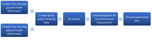
models. The scanning process was as flowing
(Figure 1a). Connected Trios correctly,
selected the research model function and entered the scan interface. The
scanner was placed on the lingual side of the middle incisor, at an angle of 45°
to the teeth. After starting the scan, the scanning gun moved slowly to the
distal of the dental arch. During the movement, it always faced the teeth at a
45° angle. After bypassing the distal surface of the second molar,
the scanning gun was slowly moved back from buccal side of the second molar to
the middle line of the dental
arch. Scan the other side
of the dental arch in the same way and control the number of scanned sheets to
400 pictures. Figure
1(a): Experimental flow
chart. After opening the 3 Shape Dental
System (3 Shape Incorporated, Denmark), we used the 3D preview function to
reconstruct the 3D version of the impression (Figure 1b), capture the 2D plane, and measured the distance of the
points on each model separately. We measured the buccal-lingual
thickness of maxillary central incisors, the length from the medial to the
distal of buccal cusps of maxillary second molars, the crown width of mandibular
central incisors, the
buccal-lingual thickness of maxillary central incisors requiring a veneer, the
buccal-lingual width of the inlay of the mandibular
first molar. Each model was
measured three times and recorded as A1, B1, C1, D1, and E1. Scanned the same model in the same
way with a slower scanning speed, increased the image stitching, and controlled
the number of scanned sheets to 1000 sheets as A2, B2, C2, D2, E2. The paired t-test was used to
compare the differences between the two groups. Data was shown in mean ± standard
deviation in each group. The test level α was set to 0.05. P <0.05 was
considered statistical significant. The
effect of scanning number on the precision of digital impression of natural
tooth The 1000 scanning number groups
caused more time while the reconstruction process of 400 scanning number groups
were more smoothly. Whats more, splicing distortion could be obviously observed
in the 1000 scanning number groups in the 3D reconstructed models. Especially,
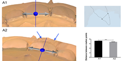
the distortions in the labial side of the crown and gingival of maxillary
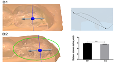
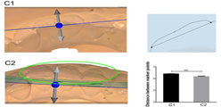
incisor in A2, the occlusal surface in B2, the labial side of the crown in C2
were severed compared with the 400 scanning number groups (A1, B1, C1) (Figures 2-4). The differences of
scanning data between the three groups were: 7.221 ± 0.010mm (A1), 6.768 ± 0.069mm
(A2). 5.839 ± 0.06mm (B1), 5.524 ± 0.025mm (B2). 4.802 ± 0.036mm (C1), 4.332 ± 0.044mm
(C2) (Supplementary file 1). They
were all statistically significant (P <0.001).
All of these suggested that 1000 scanning number required more time but it
could not offer a more smoothly scanning process and induced distortions in the
reconstruction of 3D models. Figure 2: Three-dimensional preview and two-dimensional measurement
diagram of group A. ***P <0.001. Figure 3: Three-dimensional preview and two-dimensional
measurement diagram of group B. ***P <0.001. Figure 4: Three-dimensional preview and two-dimensional
measurement diagram of group C. ***P <0.001. The
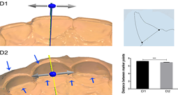
effect of scanning number on the precision of digital impression of maxillary
central incisors requiring a veneer D2 in the anterior veneer
preparation group could not offer more details of the prepared teeth, but it
caused more time in the scanning process. And the distortion could be found in
the ridge of the labial and lingual side of the crown in maxillary central
incisor (Figure 5) which could
affect the esthetic outcome and caused restoration
failure of the veneer. Whats more,
the difference of scanning data between two groups was: 7.23 ± 0.026mm (D1),
6.932 ± 0.020mm (D2) (Supplementary file
1). The difference of measurement was statistically significant (P <0.001). Figure 5: Three-dimensional preview and two-dimensional
measurement diagram of group D. ***P <0.001. The
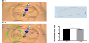
effect of scanning number on the precision of digital impression of mandibular
first molar inlay Similar to group D, E2 in inlay
tooth preparation group also didnt display more image details, but it took
longer time than group D, and the reconstruction efficiency was poor. The
distortion also could be found in the buccal side of the crown. The difference
of scanning data between the two groups was: 6.568 ± 0.022 (E1), 6.368 ± 0.023
(E2) (Supplementary file 1). The
difference of measurement was statistically significant. (P <0.001) (Figure
6). Figure 6: Three-dimensional preview and two-dimensional
measurement diagram of group E. ***P<0.001. This distortion in the 1000
scanning number groups would cause incompatibility which affected product
efficacy adversely or induced restoration failure. Theoretically speaking, more
scanning number for a model will increase the accuracy of the digital model. In
this study, we explored the appropriate parameters of scanning number for
producing the accuracy of digital impression in clinical practice. We found
that 1000 scanning number groups could not offer more details compared to 400
scanning number groups, and it could cause 3D reconstruction distortion and
longer scanning time all of which could induce esthetic out problem and restoration
failure. In 1985, Professor Werner Mormann
put forward the concept of digital impression for the first time; a new
technology of impression making based on digital idea came into being [9].
Researchers [10] studied the edge vertical distance of 30 right maxillary
second premolars made by traditional impression method and digital impression
method. The results showed that the edge adaptability of single crown by
digital impression method was better than that of traditional impression
method. The scholars compared the intraoral digital model obtained by Trios
with the intraoral model prepared by silicone rubber, and the results showed
that there was no significant difference between them. It was considered that
the accuracy of intraoral scanning digital impression can reach the clinical
standard [11]. Ultrafast Optical SectioningTM
[12] and confocal microscopy are used in the Trios system, so the scanning
speed per unit time is faster than that of other systems. Sandblasting on the
surface of teeth and soft tissues can be omitted, which greatly shortens the
operation time and improves the acceptance of patients [13]. The digital model
is displayed on the screen as a three-dimensional image. The operator can
rotate the model and observe the shape of the abutment from multiple angles
[14], and transmit clinical information to the studio simply and quickly. At
the same time, patients are more inclined to accept the way of making digital
impressions by intraoral chairside scanning [15,16]. Considering the complicated
intraoral situation, we chose the standard gypsum model, which required that
the gypsum model cannot reflect light and be easily scanned and imaged. When
using Trios, we need to determine the number of scanning sheets according to
the size of the area to be scanned. This is mainly to avoid data mosaic caused
by repeated scanning. In this experiment, the image deformation of the
three-dimensional preview was observed in the experimental groups A2, B2, and
C2, This may be due to the fact that when scanning normal natural teeth, the
number of scanned images was too large, so that the image was fitted when the
three-dimensional image was reconstructed, which caused the image to be
deformed. The thinnest veneer is 0.2mm-0.3mm, which requires an extremely high
precision [17]. In the scanned image of group D2, the cutting edge of the
preparation part was rough and not smooth, which might mislead the mechanic to
make a veneer that didnt match the preparation body, and eventually lead to
restoration failure of the veneer. Since the number of scans was increased, the
data processing time was very slow. We observed that during the scanning of the
anterior veneers and posterior inlays, the scanning time was greatly extended
and the 3D reconstruction efficiency decreased significantly. In order to avoid
data splicing during digital scanning, changing the angle to fill the unswept
area is an effective way to produce a more accurate
prosthesis and the scanning time
can be reduced. During the scanning process, when the number of scanned sheets
is about 1000, the device will be stuck, which greatly affects the scanning
efficiency. According to our experiment, if
there are too many scanning sheets in the same area, it will cause data mosaic,
and it will take more time for pictures to build three-dimensional images,
which will result in errors and data distortion, so too many scanning sheets
have an impact on the accuracy of digital impression. And further research is
needed to investigate the effect of other parameter on the clinical
scanning accuracy. Our study was supported by the
National Natural Science Foundation of China (31400808, 81570979, and
21402018). This study was also supported by Science & Technology Committee
of Yubei District (2015-01), Program for Innovation Team Building at
Institutions of Higher Education in Chongqing in 2016 (CXTDG201602006) and
Chongqing Municipal Key Laboratory of Oral Biomedical Engineering of Higher
Education (Yujiaoke (2014)-55) and the Science and Technology Innovation
Program of Social Undertakings and Peoples Livelihood Security of Chongqing
Science and Technology Commission (cstc2016shms-ztzx0045). And including this
study, Xiaomian Wu was awarded the Excellent Graduation Thesis Instructor of
Chongqing Medical University in 2018. 1.
Birnbaum NS and Aaronson
HB. Dental impressions using 3D digital scanners: virtual becomes reality
(2008) Compend Contin Educ Dent 29: 494, 496 and 498. 2.
Fasbinder DJ. Computerized
technology for restorative dentistry (2013)
Am J Dent 26: 115. 3.
Abdulaziz ABA,
Kiho K, Finkelman MD, Zandparsa R and Hirayama H. The effect of variations in
translucency and background on color differences in CAD/CAM lithium disilicate
glass ceramics (2014) J Prostho 23: 213-220. https://doi.org/10.1111/jopr.12080 4.
Davidowitz G and
Kotick PG. The Use of CAD/CAM in Dentistry (2011) Dent Clin North Am 55: 559-570.
https://doi.org/10.1016/j.cden.2011.02.011 5.
Tsitrou EA,
Northeast SE and Noort RV. Evaluation of the marginal fit of three margin
designs of resin composite crowns using CAD/CAM (2007) J Dent 35: 68-73.
https://doi.org/10.1016/j.jdent.2006.04.008 6.
Aragón ML,
Pontes LF, Bichara LM, Flores-Mir C and Normando D. Validity and reliability of
intraoral scanners compared to conventional gypsum models measurements: a
systematic review (2016) Eur J Orthod 38: 429. https://doi.org/10.1093/ejo/cjw033 7.
Amin S, Weber
HP, Finkelman M, El Rafie K, Kudara Y, et al. Digital vs. conventional
full‐arch implant impressions: a comparative study (2016) Clin Oral Implants
Res 28: 1360-1367. https://doi.org/10.1111/clr.12994 8.
Renne W, Ludlow
M, Fryml J, Schurch Z, Mennito A, et al. Evaluation of the accuracy of 7
digital scanners: An in vitro analysis based on 3-dimensional comparisons
(2017) J Prosthet Dent 118:
36-42. https://doi.org/10.1016/j.prosdent.2016.09.024 9.
Pieper R.
Digital impressions--easier than ever (2009) Int J Comput Dent 12: 47-52. 10.
Ng J, Ruse D and
Wyatt C. A comparison of the marginal fit of crowns fabricated with digital and
conventional methods (2014) J Prosthet Dent 112: 555-560. https://doi.org/10.1016/j.prosdent.2013.12.002 11.
Fleming PS,
Marinho V and Johal A. Orthodontic measurements on digital study models
compared with plaster models: a systematic review (2011) Orthod Craniofac Res
14: 1-16. https://doi.org/10.1111/j.1601-6343.2010.01503.x 12.
Berrendero S,
Salido MP, Valverde A, Ferreiroa A and Pradíes G. Influence of conventional and
digital intraoral impressions on the fit of CAD/CAM-fabricated all-ceramic
crowns (2016) Clin Oral Investig 20: 2403-2410. https://doi.org/10.1007/s00784-016-1714-6 13.
Xie YL and Shen
G. Accuracy and reproducibility of intraoral scanning in vivo (2016) Shanghai
Kou Qiang Yi Xue 25: 593-599. 14.
Sang J, Lee and
Gallucci GO. Digital vs. conventional implant impressions: efficiency outcomes
(2013) Clinical Oral Implants Research 24: 111-115. https://doi.org/10.1111/j.1600-0501.2012.02430.x 15.
Wismeijer D,
Mans R, Van GM and Reijers HA. Patients preferences when comparing analogue
implant impressions using a polyether impression material versus digital
impressions (Intraoral Scan) of dental implants (2015) Clin Oral Implants Res 25: 1113-1118. https://doi.org/10.1111/clr.12234 16.
Yuzbasioglu E,
Kurt H, Turunc R and Bilir H. Comparison of digital and conventional impression
techniques: evaluation of patients perception, treatment comfort, effectiveness
and clinical outcomes (2014) Bmc Oral Health 14: 10-10. https://doi.org/10.1186/1472-6831-14-10 17. Turgut S, Bagis B and Ayaz EA. Achieving
the desired colour in discoloured teeth, using leucite-based cad-cam laminate
Systems (2014) J Dent 42: 68-74.
https://doi.org/10.1016/j.jdent.2013.10.018 *Xiaomian Wu,
Chongqing Key Laboratory of Oral Diseases and Biomedical Sciences, College of
Stomatology, Chongqing, Medical University, Chongqing, China, E-mail: wuxiaomian@hospital.cqmu.edu.cn
, wuxiaomian898@163.com
*Ping Ji, Department
of Oral and Maxillofacial Surgery, Stomatological Hospital of Chongqing Medical
University, 426 Song-Shi North Road, Chongqing, 401147, P.R. China, E-mail:
jiping@hospital.cqmu.edu.cn Yang ZQ, Deng F, Hu XL, Wen YX, Ji P, et al. The
effect of the scanning number on accuracy of digital impression (2019)Dental Res Manag 3: 38-41 Digital impressions, Oral impression technique,
Scanning number, 3 shape trios.The Effect of the Scanning Number on Accuracy of Digital Impression
Zhi-Qiang Yang,Feng Deng,Xiao-Lei Hu, Yu-Xuan
Wen,Ping Ji and Xiao-Mian Wu
Abstract
Materials and methods: 30 standard gypsum models were divided into group
A, B, C, D and E. Each group was scanned with 400 and 1000 pictures by Trios.
Then Using the three-dimensional preview function to compare the fitted images
and to measure the distance between markers: the buccal-lingual thickness of
maxillary central incisors, the length from the medial to the distal of buccal
cusps of maxillary second molars, the crown width of mandibular central
incisors, the buccal-lingual thickness of maxillary central incisors requiring
a veneer, and the buccal-lingual width of the inlay of the mandibular first
molar.
Results: The scanning time of 1000 scanning number groups
were significantly prolonged and the efficiency of 3D reconstruction was
significantly reduced. But obvious image stitching distortion could be observed
in these groups compared with 400 scanning number groups. Whats more, the
compare of measured values in each 400 and 1000 scanning number group was all
statistically significant.
Conclusion:
The data splicing caused by the excessive scanning number may influence the
digital impression accuracy. Trios scans 400 pictures was with higher scanning
accuracy than scanning 1000 pictures. Full-Text
Introduction
Materials
and Methods

Statistical
Analysis
Results
Discussion
Acknowledgments
References
*Corresponding authors
Citation
Keywords
