The study was carried out to compare the Vickers
Micro-Hardness (VHN) of composite resin of different diameters (6 mm and 10 mm)
prepared in a split mold. The 6 mm and 10 mm diameter composite resin samples
were cured with the tip of a fiber optic light. Additional sample for same size
were cured with the same tip after a mirror or a lens accessory was mounted to
it. All the specimens were cured for 40 seconds. The hardness was calculated
for both top and bottom at the center and the periphery. The results showed
that the hardness of top surface was higher than that at the periphery. The
mean hardness value for specimens cured with the light tip was higher than the
hardness of specimens cured with their accessory. Conclusion mirror or lens
didn’t potentiate light but distribute the energy to a larger area, resulting
in less energy applied to the same small surface area, which reduced the
hardness.
Introduction
Visible
light-cured resin systems have expanded the
versatility of composite resin in dentistry. Since many large restoration
composites such as veneers are being placed. Adequate
polymerization of the entire restoration is a major concern. Also, it is very important
for the dentist to cure restorations in the minimum time possible to maximize
office productivity without compromising the long term success of the
restorations [1].
A
major disadvantage of the available light generating units is the small
diameter of light tip, typically 5-7 mm which results in small area of cure.
Curing the entire restoration at one time instead of increments would help to
solve lamination problems, and reduce the time needed to restore an esthetics
veneer. Light cure a large area of composite
restoration by scanning the composite resin surface
results in a cure which is lower in hardness than cured with fixed
illumination. A small tip diameter may deliver a high radiant existence;
multiple exposures may be required to completely cover the restoration [2-4].
The
purpose of this research was to compare the micro-hardness of composite
surfaces of 6 and 10 mm diameter specimens cured after a large diameter a
highly polished metal ring (3M company, USA, St, Paul) (Figure 1) that collect all the scattered rays from the light tip,
expected to increase the energy to cure composite resin, or the (Kulzer Translux
CL photo cure attachment lens) (Figure 2).
Which will focus the rays in the center and concentrate it to increase the
potency of the rays expect more composite cure were mounted, to a light unit
rod to those cured with the light cure unit rod without accessories.
The
null
hypothesis is that, resin composite light curing
unit with the accessories give the same test of Vickers hardness as without
when curing large diameter composite.
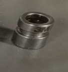
Figure 1: Mirror, a highly polished metal ring.
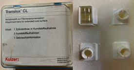
Figure 2: Kulzer Translux CL photo cure attachment lens was attached.
Material and Methods
A
universal color of microfilmed composite (Helio Progress) was used, Thirty
specimens 10 mm in diameter and 2 mm thick and thirty specimens of 6 mm in
diameter and 2 mm thick were prepared in a split mold. The split mold was
placed between two Mylar strips and covered with a glass slide to assure a
smooth surface which could be a measured accurately. The mold cavity was
overfilled with composite and pressed against the Mylar strip and glass slide
to remove the excess. Ten specimens of 6 mm diameter (Group I) and ten of 10 mm
diameter (Group IV) were exposed for 40 seconds to light from a light cure unit
(Vislux, 3M Company, USA, St. Paul). Ten specimens of composite with 6 mm
diameter (Group II) and ten specimens of 10 mm diameter (Group V) were exposed
to light form the same light cure unite for 40 seconds, after the mirror, a
highly polished metal ring was attached. Ten additional specimens of composite
with 6 mm diameter (Group III) and ten specimens of 10 mm diameter (Group VI)
were exposed to light from the same light cure unit, after was attached. kulzer
and CO. GMBH, wehrheim-Germany) (Figure
3) a box of four frames to be used alternatively during sterilization while
the lens attached to one frame.

Figure 3: 3M lens attached to the lens.
All
the three groups were subdivided according to the surface measured. The top
surface of the 6 mm specimens was labeled “a”, and the bottom surface was
labeled “b” similarly the top of the 10 mm specimens were labeled “c” and the
bottom “d”. The tip of the fiber optic light unit alone and with added accessories
mirror or with lens were placed in contact with the glass slide over the mold
to cure the corresponding group of the three composite
resin groups.
All
the cured specimens were kept in a light proof container until the hardness
test was performed. The vicker’s
micro-hardness test was performed. Micro-hardness tester
(Vickers Shimadzu-Seisa Kusho Ltd. Micro-hardness Tester,Kyoto, Japan), by measuring
three indentations left by 100 gm sustained for 16 seconds. A diamond shaped
indenting needle was used and each diagonal was measured. The mean value was
calculated, that correlate the hardness with mean of diagonals obtained at the
center and periphery.
Results
The
mean VHN hardness value of 6 mm diameter for the three groups is shown in Table 1. A multi factorial ANOVA
performed for comparison of hardness of the three groups at 1-a, 2-a, 1-b and
2-d areas (F = 33.10, P<0.05). The Duncan test shows that the mean hardness
value for group I is significantly higher than that for group II and III. The
mean hardness values of 10 mm diameter for the three groups are shown in Table 2. A multi factorial ANOVA was
performed for comparison of hardness of three groups at 1-c, 2-c, 1-d, and 2-d
areas (f = 33.10, p<0.05). The Duncan test shows that the mean for group I
and II is significantly higher than group III and the difference between that I
and II is not significant. Hardness comparison of 6 and 10mm diameter of the
six groups by performing a multi factorial ANOVA showed that there are
significant differences in values between individual cases (F = 48.86,
P<0.05) (Table 3). The Duncan
test shows that the mean for Group I is
significantly higher than that for all groups. Similarly, the mean for hardness
values of Groups II, IV, and V are significantly higher than that of Groups III
and VI. However, there are no significant differences between the mean hardness
values of Groups II, IV, and V. There are no significant differences between
the mean hardness values of Groups III and VI.
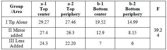
Table 1: The mean Vickers hardness value for the three group of 6 mm diameter.
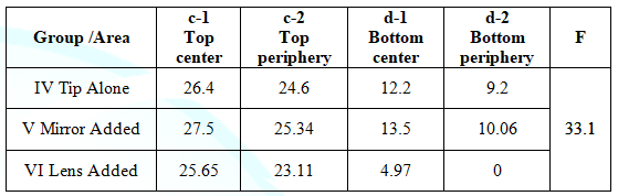
Table 2: The mean Vickers hardness value for the three group of 10 mm diameter.
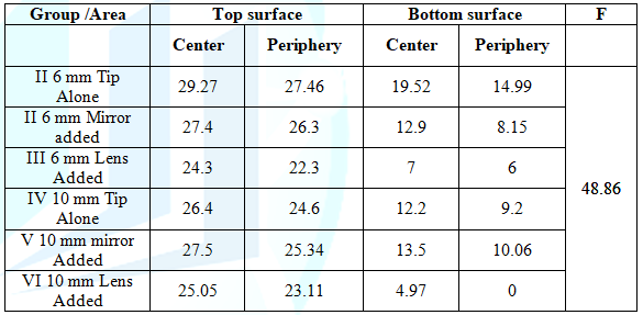
Table 3: Comparison of the six groups of 6 and 10 mm Diameter.
Discussion
The
hardness of all 6 and 10 mm specimens shows that the hardness of 6mm specimen
cured with the light unit rod showed the highest of all. This might be because
direct light shows the maximum effect especially when direct contact with the
specimens and is without Intervening accessories this agreed with Leonard et al
[5] who found that fixed and direct contact of small light tip with is
essential to perform good composite resin cure.
Addition
of accessories to the unit tips although it is try to collect the scattered
light waves and concentrate at the center it but showed reduced in cure as
indicated by the resulted hardness obtained this might be due to that the light
tips away by distance from direct contact with composite resin this agreed with
Gabriel et al [6] who stated that increase the distance of light tip by more
than 5mm will affect the depth of cure of composite resin material. And S Zhu
and J Platt [7] who found that increased the distance inversely proportion with
the distance.
The
results of this study showed descending gradation in hardness in all the
specimens tested, from center to the periphery, from top to bottom, and from
small light curing unit rod to a larger (added accessories), this may because
light from the curing unit is a sort of the energy in a cone form concentrated
at the center helping to activate inactive molecules. that the light generation
produced a conical pattern of hardness at the center and gradually drop off
toward the periphery of the tip increasing the polymerization, decreasing the
conical effect of polymerization with the confined diameter of the light tip
local hardness values of the specimens are plotted as hardness maps, this
agreed with Pires, et al [8] who stated that the hardness decreased from center
to periphery and from top to bottom. Also Price and McLeod [9] who stated that
light is a sort of energy that decreased away from the light curing tip margins
or distance.
Our
results showed that when accessory with larger diameter are added to the light
unit of 6 mm diameter, a decrease in Vickers hardness of composite resin. The
lens shows least hardness of all. This may be due to the fact that the light
cure unit emits light with the same energy for all the Specimens that reduced
as it passed through the lens and its thickness that increases the distance to
the specimens. This agreed with Sobrinho CL et al [10] agreed that The use of
wide diameter curing light tip from outside the cavity may result in incomplete
curing.
Our
results showed that 10 mm diameter specimens that were cured with attached
lens, has a significant decrease in hardness to the extent that its
measurements couldn’t be performed. This suggests that light energy is reduced
as it passed through the lens it was not enough to cure the bottom surface.
This agreed with Thomé T et al [11] who agreed that the degree of
polymerization of light activated composite increased with direct and more
exposure to the photo activating light. Also Kwon PC and Park WJ [12] who
stated that the top surface of cured composite resin is harder than the bottom.
Conclusion
- Within
the limit of this study it is concluded that:
- Maximum composite resin cure
which is essential to obtain successful restorative
material was demonstrated by its increased in
hardness.
- Light is sort of energy which
helps to cure of composite resin and gets higher hardness allow energy
concentrated at the smaller tip than larger ones at its center.
- The hardness of resin composite
at the center of top surface and focused light was higher than that at the
periphery of the light unit tip.
- Curing of composite restorative
material showed hardness of the top surface faced light curing tip at both
center and periphery were higher than the bottom surface away from it whether
at bottom center or periphery in all the composite resin specimens.
- Curing of larger diameter of
composite resin than the curing light tip showed reduced hardness than smaller
diameter of composite resin as indicated by its hardness when not fixed the
light unit tip on the field of cure.
- The mean hardness value for
specimens cured with light tip was higher than the hardness of specimens cured
with both added accessories trying to reflect the scattered
rays or by focus it on the composite
resin specimens.
References
- Mousavinasab
SM and Meyers L. Comparison of depth of cure, hardness and heat generation of
LED and High intensity QTH light sources (2011) Eur J Dent 5: 299-304. https://doi.org/10.1055/s-0039-1698895
- Price
RB, Murphy DG and Derand T. Light energy transmission through cured resin
composite and human dentin (2000) Quintessence Int 31: 659-667.
- Nitta
K. Effect of light guide tip diameter of LED-light curing unit on
polymerization of light-cured composites (2005) Dent Mater 21: 217-223. https://doi.org/10.1016/j.dental.2004.03.008
- Haenel
T, Hausnerová B, Steinhaus J, Price RBT, Sullivan B, et al. Effect of the
irradiance distribution from light curing units on the local micro-hardness of
the surface of dental resins (2014) Dental Materials 31: 93-104. https://doi.org/10.1016/j.dental.2014.11.003
- Leonard
DL, Charlton DG and Hilton TJ. Effect of curing-tip diameter on the accuracy of
dental radiometers (1999) Oper Dent 24: 31-37.
- Corciolani
G. Vichi A, Davidso LL and Ferrari M. The influence of tip geometry and
distance on light-curing efficacy (2008) Oper Dent 33: 325-331. https://doi.org/10.2341/07-94
- Zhu
S and Platt J. Curing Efficiency of three different curing modes at different
distances for four composites (2011) Oper Dent 36: 362-371. https://doi.org/10.2341/09-245-l
- Pires
JA, Cvitko E, Denehy GE and Swift EJ. Effects of curing tip distance on light
intensity and composite resin microhardness (1993) Quintessence Int 24: 517.
- Price
RB, McLeod ME and Felix CM. Quantifying light energy delivered to a Class I
restoration (2010) J Can Dent Assoc 76: a23.
- Sobrinho
C, Lima A, Consani S, Sinhoreti CM and Knowles CJ. Influence of curing tip
distance on composite knoop hardness values (2000) Braz Dent J 11: 11-17.
- Thomé
T, Steagall W, Tachibana A, Braga SR and Turbino ML. Influence of the distance
of the curing light source and composite shade on hardness of two composites
(2007) J App Oral Sci 15: 486-491. https://doi.org/10.1590/s1678-77572007000600006
-
Kwon PC and Park WJ. Influence of thickness on
the degree of cure of composite resin core material (2006) J Korean Acad
Conserv Dent 31: 352-358. https://doi.org/10.5395/jkacd.2006.31.5.352
*Corresponding author
Samir Koheil, Professor, Department of conservative
dentistry and implant, Faculty of Dentistry, Alexandria University, Egypt,
E-mail: skoheil@yahoo.com
Citation
Koheil S. A study of composite
surface hardness when cured using special accessories with a curing light
(2020) Dental Res Manag 4: 11-13.
Vickers micro-hardness, Composite resin, Dentistry.


