Lipoid pneumonia which is a rare disorder, results from accumulation of lipids in the alveoli. Lipoid pneumonia has two types; exogenous pneumonia which results from deposition of inhaled oil in the lung and endogenous pneumonia which results from breakdown of lung distal to obstructed airway. Tumor or bronchiolitis obliterans or following chemotherapy or radiotherapy lead to release of cholesterol and other lipids in alveoli. We report clinical and radiological findings of two cases with exogenous lipoid pneumonia due to aspiration of oil products.
Introduction
Lipoid
pneumonia which is an uncommon disease, with a frequency of 1-2.5% caused
by the presence of lipid in the alveoli [1,2]. Lipoid pneumonia is classified
into two major groups; exogenous and endogenous/idiopathic pneumonia [2]. Acute
exogenous pneumonia is typically caused by aspiration of a sum of
petroleum-based products [2]. It is typical for children due to accidental
poisoning [2]. Chronic exogenous pneumonia commonly results from recurrent
episodes of animal fat, mineral or vegetable oils [2]. Exogenous pneumonia is
typically for older patients but sometimes it can be seen in children suffering
from aspiration
[2]. Lipoid pneumonia commonly presents with subtle onset and respiratory
symptoms such as cough, dyspnea,
chest pain, fever, hemoptysis, systemic findings and radiological findings such
as pulmonary opacities, consolidations, pneumomediastinum, pneumothorax,
pleural effusions, pneumotoceles and nodules [1,2]. We aim to present here two
cases of exogenous pneumonia and their radiological findings.
Case Report 1
A 16-year-old boy admitted to
emergency room (ER) with nausea, emesis, cough, sputum and chest pain. His
medical history and family history were unremarkable. He said that he took
diesel oil with a hose by mouth from the chemical reservoir and he aspirated it
accidently one day before. The patients general condition was worsened during the
examination
and he was intubated in ER. In the respiratory system examination; inspiratory
rales were heard more prominent in the on the right lung basal side. Figure 1a-1c show chest
radiographs of him on the admission, 10th day and 30th day, respectively.
The patient was given antibiotics (Meropenem 1 gram, three times a day) in the
intensive care unit. The patient was extubated and taken to Respiratory
medicine service on the 4th day of the treatment. Verbal consent was obtained
from the patient for this case presentation.
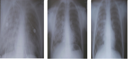
Figure 1a-1c: Chest radiographs of him on the admission, 10th
day and 30th day, respectively.
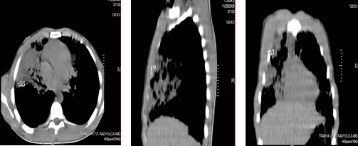
Chest computed tomography showed
parenchymal
consolidation areas containing air bronchograms at fat density. Density
measurements were made in vertical, axial and sagittal sections.
Figure 1d-1f show chest computed
tomography findings of exogenous pneumonia for this patient.
Antibiotherapy of the patient continued with ampicillin-sulbactam (4*1.5 gram
intravenous for a day) and completed to 15 days. The patient with clinical and
radiological improvement was discharged at the end of 15 days. After discharge,
nodular sequela was seen on the patients chest tomography (Figure 1g and 1h).
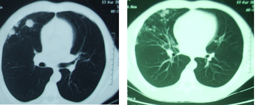
Figure 1g and 1h: Noduler sequela was
seen on the patients chest tomography in the sixth month, after discharge.
Case Report 2
A 72-year-old man admitted to
emergency room with cough, high fever and hemoptysis. The patient accidentally
drank a glass of gas oil five days before. The patient had only hypertension
disease. 4 hours after the incident, the patient was admitted to a hospital
and treated with antibiotics.
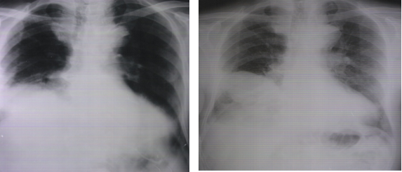
Figure 2a and 2b: Chest radiographs of the patients admission to
hospital and hospital discharge.
The patient was referred to our
hospital due to persistent high fever and hemoptysis. The patient was
conscious, cooperative, and poor in orientation. In the
respiratory
system examination, breathing sounds were reduced in both lungs and there
were inspiratory rales in the right middle and lower left lungs.
Bronchoscopy
revealed no endobronchial lesion. Mucosa of right middle and lower lobes were
with edema. Cytology showed no lipid-laden macrophage. Ampicillin-resistant
E. coli and
C. Albicans were seen as a result of
bronchoalveolar
lavage. The patients antibiotic treatment was continued with cefuroxime
sodium and ciprofloxacin. The thoracentesis performed from the patients pleural
effusion was consistent with exudates.
Oral
antibiotic was initiated on the 7th day. The patient was discharged on the
14th day of treatment. Verbal consent was obtained from the patient for this
case presentation.
Discussion
The exogenous lipoid pneumonia is
caused by aspiration or inhalation of mineral oils, animal oils or petroleum
products or lipoid pneumonia can be endogenous commonly following post
obstruction either due to lung cancers, bronchiolitis or lung necrosis [3]. One
of the our patients who presented with lipoid exogenous pneumonia was diesel
oil aspiration and the other one was admitted to ER as a result of gas oil
aspiration.
In the literature, case reports
related to aspiration-induced lipoid pneumonias were reported in childhood.
However, lipoid pneumonia due to oil aspiration can also be seen in elderly
patients. Our patient who was exposed to gas oil aspiration was 72-years-old
man. The clinical symptoms of lipoid pneumonia are nonspecific and may vary due
to patients age, duration of oil intake; the amount and quality of oil
aspirated [4]. In the literature, time of exposure, amounts of oils and quality
of material vary according to case reports. Elder patients are more
asymptomatic than early ages and also lipoid pneumonias are commonly chronic
and progressive for geriatric
patients [4]. Because of this reason, lipoid pneumonias are usually discovered
by as an autopsy finding [4]. Both of our patients presented to the emergency
department with severe clinical presentation.
The diagnosis of exogenous lipoid
pneumonia is based on a history of exposure to oils and clinical findings,
radiological findings, presence of lipid-laden macrophages on sputum
or bronchoalveolar lavage [4]. In despite of, chest radiographs may be
unremarkable in patients with exogenous lipoid pneumonia; most of them show
abnormalities [5]. Homogenous dense consolidation with air bronchograms,
diffuse or focal and unilateral or bilateral involvement, cavitation, nodules
and masses resulting from inflammation or fibrosis, atelectasis and pleural
effusions may seen on chest radiographs of these patients [5]. Chest computed
tomography and magnetic resonance imaging may detect fats within pulmonary
tissues [5]. However, that none of these clinical and radiological findings
alone is diagnostic for exogenous
lipoid pneumonia.
Treatment modalities of lipoid
pneumonia are poorly defined. Treatment strategies may include whole lung
lavage, supportive care, systemic corticosteroids
and thoracoscopy with surgical debridemen [6]. Antibiotics treatment may be
given for complicated patients. Lipoid pneumonia is an uncommon disease
encountered in all age groups. Physician should enquire about oil intake in all
patients with persistent cough and chest symptoms. Treatment involves removal
of the offending agent and supportive care.
References
1. Hadda V and Khilnani GC. Lipoid
pneumonia: An overview (2010) Expert Rev Respir Med 4: 799-807. https://doi.org/10.1586/ers.10.74
2. Betancourt SL, Martinez-Jimenez
S, Rossi SE, Troug MT, Carrillo J, et al. Lipoid pneumonia: Spectrum of clinical
and radiologic manifestations (2010) Cardiopulmonary Imaging 194: 103-105. https://doi.org/10.2214/AJR.09.3040
3. Sharma A, Ohri S, Bambery P and
Singh S. Idiopathic endogenous lipoid pneumonia (2006) Indian J Chest Dis
Allied Sci 48: 143-145. https://doi.org/10.1016/j.rmed.2010.12.001
4. Marchiori E, Zanetti G, Mano CM
and Hochhegger B. Exogenous lipoid pneumonia. Clin radiological manifestations
(2011) Respiratory Med 105: 659-66. https://doi.org/10.1016/j.rmed.2010.12.001
5. Banjar H. Lipoid Pneumonia: A
review (2003) Bahrain Medical Bulletin 25: 1-6.
6. Shaikh A and Oliveire PJ.
Exogenous lipoid pneumonia (Fire-eaters Lung) (2014) American J Med 127: 3-4. https://doi.org/10.1016/j.amjmed.2013.10.008
*Corresponding author: Sema Avci,
Emergency
Medicine, Amasya University, Sabuncuoglu Serefeddin Research and Training
Hospital, Turkey, E-mail: dnzlsema@gmail.com
Citation: Perincek G, Avci
S and Batmaz E. Lipoid pneumonia due to aspiration of
oil products: Two case reports (2018) Nursing and Health Care 3: 69- 70
Lipoid pneumonia, Aspiration, Oil products