Congenital hemifacial
hyperplasia (CHFH) is a rare congenital malformation characterized by marked
unilateral overdevelopment of the hard and soft tissues of the head and face
region. The exact etiology is not clear it is usually evident at birth and increases
with age, mostly till puberty. In some cases multiple systems are involved. Treatment
is usually indicated in cases where esthetics is a concern.
A 6 year old female reported to the Department
of Oral Medicine and Radiology with the features of congenital hemifacial
hyperplasia, we are making an attempt to present a rare case of CHFH in order
to highlight the clinical manifestations and treatment options.
Introduction
Hemifacial hyperplasia or
hypertrophy is a condition in which one half of the face either maxilla or
mandible alone or soft tissues of one half of the face grows to unusual proportion
as compared to the opposite side. It is a rare congenital malformation. This condition
has been termed as facial hemihyperplasia, partial / unilateral gigantism, hemimacrosomia, or hemifacial
hypertrophy [1].
CHFH was first noted by Meckel JF
in 1822, as a developmental anomaly and the first case was reported by Wagner
in 1839[2]. Hyperplasia can be seen in association with several syndroms like
Beckwith Weidemann syndrome, Proteus syndrome, Russell silver syndrome and Sotos
syndrome. Gessel in 1927 described CHFH as essentially a developmental anomaly
antedating birth. Rowe[3] in 1962 proposed a classification of hemifacial
hyperplasia based on its anatomical location as simple (one or both limbs), complex
(entire half of the body), and facial hemihyperplasia( face, head and associated
structures). Based on soft tissue involvement HFH can be classified as true (increased
growth of not only the soft tissues of the face but the hard tissues as well) and
partial (increased growth limited to one structure only) [4,5].
We present an interesting case of
a 6 year old female with a true congenital hemifacial hyperplasia.
Case
Report
A 6 year old female patient
reported to the Department of Oral Medicine and Radiology with the chief
complaint of a large upper left front tooth since six months. Patient’s mother
was concerned about the esthetics. They had noticed an enlargement of the left side
of the face since birth, which has been progressing in size. Patient had an
early exfoliation of the upper left deciduous tooth which was followed by an
early eruption large sized permanent tooth. The child was the second of two
siblings born of a non-consanguineous marriage. There was no significant
prenatal or relevant medical history. Milestones were normal and she has normal
intelligence. Patient was moderately built and nourished with normal gait. The
vitals were within normal limits.
Extraoral examination revealed an
obvious swelling of the left upper half of the face, leading to asymmetry
extending superiorly from the infra orbital rim to the left upper lip inferiorly.
Medially extending from the bridge of the nose, obliterating the nasolabial
fold and laterally 2 cm anterior to the pretragal area on the left side. The
left half of the upper lip was hypertrophied and the angle of the mouth on the
left side was tilted downwards lip was incompetent. The overlying skin appeared
normal. On palpation the swelling was soft in consistency, non-tender, with no
rise in temperature (Figure 1).
Intraoral examination revealed
premature eruption of 23, 24, 27 with macrodontia of 23. Grossly decayed 54, 65,
74, 85 and 26. Dentinal caries in 53, 62, 75, deep carious lesion in 84, rest
of the dentition was normal with age. Thickened and enlarged alveolar ridge extending
from 23 till 27. Mandible was normal. No midline shift was noticed. Buccal mucosa
and tongue appeared normal (Figures 2,3).
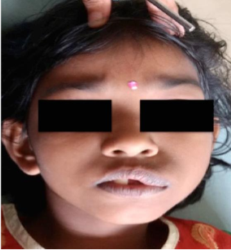
Figure 1: Extraoral Photograph.
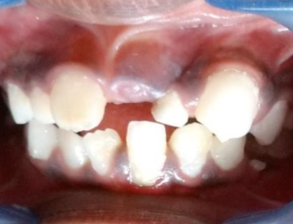
Figure 2: Macrodontia of 13.
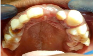
Figure 3: Enlarged left alveolar ridge.
Considering the history and
clinical features a provisional diagnosis of a congenital true hemifacial
hyperplasia of the left side was made, and a differential diagnosis of segmental
odontomaxillary dysplasia was given.
An orthopantomogram showed early
exfoliation of 63, 64 followed by early eruption of 23, 24 and 27. Enlarged
pulp chamber of 23, 24 and 27. Incomplete root formation in 23, 24 and 27. 65
also shows resorbed roots and half the root formation of 25 as compared to the
opposite side where only the crown formation is complete. Slanted palatal drag
suggestive of hyperplastic maxilla and hyperplastic zygomatic bone on the affected
side. The left maxillary sinus appears to be enlarged as compared to the right side.
There was no obvious enlargement of the left mandible including coronoid and
the condylar process (Figure 4).
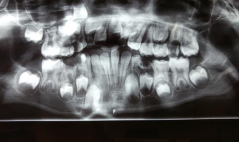
Figure 4: Panoramic radiograph.
Both the clinical and
radiographic features suggested congenital true hemi-hyperplasia on the left
side.
Patient was then referred to the
department of pedodontics for detailed examination and management. They advised
full mouth oral prophylaxis, extraction of all the teeth with grossly
destructed crown structure which includes extraction of 54, 74, 84, 85 followed
by placement of space maintainer. Since the prognosis of 26 was poor they
advised extraction of 26 also. Pit and fissure sealants and periodic follow up
was also advised. Since radiograph shows unerupted 21 and 22 due to lack of
space patient was advised orthodontic treatment followed by esthetic
recontouring of 23. Once the growth spurts are over patient was advised non-surgical
management for the facial asymmetry.
Discussion
CHFH is a rare developmental
anomaly characterized by an increased growth of both hard and soft tissues of
the face present since birth. The prevalence of HFH is approximately 1 in 86000
births [2]. In some cases it is associated with the syndromes of head and neck
region. It usually increases with age till puberty. It is usually seen more commonly
among females as compared to males mostly on the right side of the face [6]. Many
theories have been proposed as a cause of HFH including hormonal imbalances, diseases
involving the neural system, other conditions like hemangiomas, lymphangiomas, incomplete
twinning, abnormal intrauterine development, somatic mutations, mechanical
influences and congenital syphilis [7]. Numerous clinical findings may be seen
like gross asymmetry of the facial structures and presence of hypertrophied
areas. The soft tissues usually have the same consistency as that of the normal
tissues. In some cases unilateral macroglossia and prominent fungiform papillae
are also seen.
Permanent canine, bicuspids, and
first molars are most commonly involved. Premature exfoliation and early
eruption of the teeth of the affected side is noticed. In most cases teeth have
large crown size(macrodontia) and large root with
root resorption. Midline shift and malocclusion with deviated occlusal plane is
also noted. Involvement of the upper lip causes displacement of the philtrum [8,9].
Palate can also show an arch shaped deformity of the affected side. Differential diagnosis includes fibrous dysplasia,
dyschondroplasia, hemangioma,lymphangioma, A-V aneurysms, congenital
lymph edema and odontomaxillary dysplasia[10]. HFH may be associated with
skeletal deformities like polydactyly, syndactyly, scoliosis and macrodactyly. CNS
defects like epilepsy, mental retardation affects 20% of the affected patients.
HFH may also be associated with adrenal cortical carcinoma, Wilms tumor and
hepatoblastoma [11].
Treatment
Options
A multidisciplinary approach is
needed in most of the cases, includes subtle soft tissue contouring to
extensive surgeries to correct the soft and hard tissue defects. Like condylar
recontouring, osteotomies followed by debulking of the soft tissues [12]. Khanna
and Andrade did hard and soft tissue debulking to achieve acceptable results[13].
Pollock also did osteotomies and treatment of soft tissue deformities like cheiloplasty
[14]. Occlusal irregularities were corrected by orthodontic therapy. Due to the
difficulties encountered in the treatment like aggravating growth, suboptimal post-operative
esthetics has dissuaded many surgeons from approaching the condition in an aggressive
manner. Liposuction the tumescent technique has been widely used which was
introduced by Klein [15]. The other technique includes ultrasound assisted liposuction,
powered liposuction. Phosphatidylcholine preparations have been widely used for
localized reduction of subcutaneous fat. Once injected into the site the
reaction will remain for 8 to 10 weeks [16]. Despite temporary discomfort
Phosphatidylcholine injections can be successfully used in the treatment of localized
fatty areas of the face as it is safer than liposuction.
Dental abnormalities like
macrodontia, premature exfoliation, premature eruption and malocclusion can be
managed according to the abnormalities. Macrodontia with open apex can be
treated once the root completion is over either by coronoplasty or crown placement.
Premature exfoliation leading to closure of space can be managed with space
maintainers. Malocclusion can be orthodontically corrected. Carious formation due
to the premature eruption can be treated prophylatically by pit and fissure
sealants and fluoride applications.
Conclusion
To conclude this article
highlights the importance of a thorough case history, clinical examination and
diagnostic evaluation in oral medicine for the proper diagnosis and treatment
planning of such congenital developmental disturbances. Modern technology has
revolutionized treatment options, in rare conditions like this the possibility
of achieving facial symmetry is not an easy task, and it has to be done with a multidisciplinary
approach. Non-surgical management is considered a better treatment option as
compared to cosmetic surgeries.
References
1. Pollock
RA, Newmann MH, Burdi AR and Condit DP. Congenital hemi facial
hyperplasia: An embryological hypothesis and Case Report (1985) Cleft
Palate Journal 22: 173-184.
2. Mark S, Clark OH and Kaplan RA. A
virilised patient with congenital hemihypertrophy (1994) Postgraduate
Medical Journal 70: 752-755.
3. Rowe NH. Hemi facial hypertrophy:
Review of literature and addition of four cases (1962) Oral surgery,
Oral Medicine, Oral Pathology 15: 572-587.
https://doi.org/10.1016/0030-4220(62)90177-9
4. Bhuta BA.
Clinical and imaging findings of true hemifacial hyperplasia (2013) Case
Reports in Dentistry 7. https://doi.org/10.1155/2013/152528
5.
Islam M. Comparision between true and partial hemifacial hyperfacial
hypertrophy (2007) Oral Surg Oral Med Oral Pathol Oral Radiol Endod 104:
501-509. https://doi.org/10.1016/j.tripleo.2006.11.053
6.
Rodolph CE and Norvold RW. Congenital partial hemi hyperplasia involving
marked malocclusion (1944) Dent Res 23: 133.
https://doi.org/10.1177/00220345440230020501
7. Siponen M, Sandor
G, Ylikontiola L, Salo T and Tuominen H. Multiple orofacial intra
neural perineuriomas in a patient with hemifacial hyperplasia (2007)
Oral Surg Oral Med Oral Pathol Oral Radiol Endod 104: 38-44.
https://doi.org/10.1016/j.tripleo.2006.12.030
8. Lo HS. Congenital hemifacial hypertrophy (1982) British Dental Journal 153: 111-112.
9.
Verma P, Gupta K, Rishi S, Trivedi A and Kailasm S. Hemifacial
hypertrophy: A rare case report (2012) Journal of Academy of Oral
Medicine and Radiology. 24: 334-337.
10. Shanmugasundaram K, Vedam
VK, Ganapathy S, Sathish S, and Satti P. Congenital hemifacial
hyperplasia: Clinical presentation and Literature review (2016) Case Rep
Dent 5260645. http://dx.doi.org/10.1155/2016/5260645
11. Batsaki
JG. Tumors of the head and neck: Clinical and pathological
consideration (1982) William and Wilkins (2nd edtn) Baltimore 301-302.
12.
Nandimath SA, Rajkumar GC, Nayak T, Ashwin DP, Rudresh KB, et al.
Hemifacial hypertrophy: Exploring new avenues of treatment modalities
(2016) Natl J Maxillofac Surg 7: 100-104.
https://dx.doi.org/10.4103%2F0975-5950.196123
13. Pollock RA,
Newman MH, Burdi AR and Condit DP. Congenital hemifacial hyperplasia: An
embryologic hypothesis and case report (1985) Cleft palate J 22:
173-184.
14. Hall HD. An improved method for treatment of facial
asymmetry secondary to jaw deformity (1984) J Oral Maxillofac Surg 42:
673-679. https://doi.org/10.1016/0278-2391(84)90211-8
15. Klein
JA. Tumescent technique for local anaesthesia improves safety in large
volume liposuction (1993) Plast Reconstr Surg 92: 1085-1098.
16.
Palmer M, Curran J and Bowler P. Clinical experience and safety using
phosphatidycholine injections for the localized reduction of
subcutaneous fat: A multicentre, retrospective UK study (2006) J Cosmet
Dermatol 5: 218-260. https://doi.org/10.1111/j.1473-2165.2006.00257.x
Corresponding
author
Anuna Laila Mathew, Department of Oral
Medicine and Radiology, Pushpagiri College of Dental Sciences, perumthuruthy, Kerala,
India, Tel: 8547431225, E-Mail: drmathewdan@yahoo.co.in
Citation
Anuna
LM, Renu M, Gibi S and Joe J. True Hemifacial Hyperplasia-Clinical
Presentations and Treatment Options (2017) Dental Res Manag 2: 52-54


