Zimmerman-Laband
syndrome was reported by Zimmerman in the year 1928 which is a rare inherited
autosomal dominant disease characterized by generalized enlargement of the
attached and marginal gingiva, abnormalities of nose, ear, deformities of
nails, joint hyperextensibility, hepatosplenomegaly, skeletal abnormalities and
occasional mental retardation. Idiopathic gingival enlargement is usually
evident after the eruption of the permanent teeth. Both sexes are equally
affected. Genetic loci for autosomal dominant modes of gingival fibromatosis is
localized to chromosome 2p21p22 (HGF-1) and chromosome 5q12-q22 (HGF-2). This
syndrome is not a life threatening disorder. Hereditary gingival enlargement is
associated with syndromes like Rutherford syndrome, Zimmerman-Laband syndrome,
Murray-Puretic-Drescher syndrome, Cross syndrome and Ramons syndrome. The most
important feature of this syndrome is gingival enlargement appearing early in
childhood. Idiopathic gingival enlargement is usually evident after the eruption
of the permanent teeth. Surgical correction of gingival fibromatosis is
recommended, although there is no information on the permanence of the results
of this treatment. We present a case of a 14 year old female patient with
Zimmerman-Laband syndrome. Gingivectomy was carried out in the upper and lower
anterior region there by exposing the impacted teeth.
Introduction
Hereditary gingival
enlargement is associated with syndromes like
Rutherford syndrome, Zimmerman-Laband
syndrome, Murray-Puretic-Drescher syndrome, Cross
syndrome and Ramons syndrome [1].
Zimmerman-Laband syndrome is a rare entity characterized by generalized
enlargement of the attached and marginal gingiva, abnormalities of nose, ear and
deformities of nails, joint hyperextensibility, hepatosplenomegaly, skeletal
abnormalities and occasional mental retardation [2]. The prevalence of this
syndrome is 1 in 10000000. Forty four patients have been reported till date.
This syndrome has highly variable clinical expressions. Gingival
fibromatosis is characterized by slow and
progressive enlargement of maxillary and mandibular gingiva. It can be
presented as an isolated feature or as a part of syndrome. This syndrome is not
a life threatening disorder.
Etiology
The genetic basis is unknown. Mapping of breakpoints of two translocations, t
(3, 8) and t (3, 7) found in two patients with typical clinical features of
Zimmermann-Laband syndrome defined a common breakpoint region located in 3p14.3
but the lack of a specific coding-sequence
lesion in the common region suggests that
either some other type
of genetic defect in this vicinity, or an
alteration elsewhere in the genome, could be responsible for ZLS. Autosomal
dominant inheritance has been suggested.
The most important feature of
this syndrome is gingival enlargement appearing early in childhood. Idiopathic
gingival enlargement is usually evident after the eruption of the permanent
teeth [3]. The differential diagnosis includes other defined syndromes of hirsutism
and coarsening of the face. Isolated gingival fibromatosis has been documented
as a dominantly transmissible trait. Surgical correction of gingival
fibromatosis is recommended, although there is no information on the permanence
of the results of this treatment, surgical removal of the hyperplastic fibrous
tissue and appropriate orthodontic treatment to improve esthetic appearance and
eruption of the non-erupted
teeth. Our case reports a 14 year old female
patient with Zimmerman-Lanband syndrome the oral manifestation and treatment.
Case
Report
A 14 year old female patient
reported the Department of Oral
Medicine and Radiology with the chief complaint of
difficulty in chewing, enlarged gums and also about the un-erupted front teeth,
inability to close the lips causing esthetic concern. Patients parents reported
that the gingival growth gradually increased in size to the present condition.
History of recurrent cellulitis of leg was reported.
Family history revealed that her
elder brother is also suffering with the same condition hepatic
fibrosis with portal hypertension. Her parents
were normal with no features of this syndrome. Patient had normal intelligence
and is doing well in her studies. On general examination well was moderately
built and nourished with normal vital signs. Systemic evaluation revealed no
obvious internal
organ abnormalities.
On extra oral examination showed
the presence of thick eyebrows, bulbous nose and floppy ears, thick and
incompetent lips as shown in Figure 1.
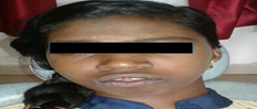
Figure 1: Extraoral photograph
Patient also had plantar
keratosis shown in Figure 2.
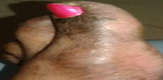
Figure
2: Plantar keratosis.
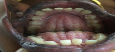
Intraoral examination revealed severe gingival
enlargement of the maxillary and mandibular anterior region with high arched
palate Figures 3, 4.
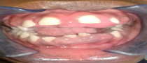
Figure
3: Intraoral photograph showing gingival enlargement.
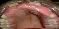
Figure
4: Showing high arched palate.
Due to the gingival enlargement
the upper and lower anterior teeth were embedded in the gingiva with partially
erupted upper and lower central incisors. Gingiva was firm, leathery in
consistency with lobulated and pebbled surface. There was melanosis of the buccal
and labial mucosa. A panoramic radiograph was
taken revealed the presence of all the permanent teeth arranged irregularly in
the anterior region Figure 5.

Figure
5: Panoramic Radiograph.
Hematological and biochemical
investigations were within the normal limits. Ultrasound revealed no signs of hepatosplenomegaly.
Patient was diagnosed with Zimmerman-Laban syndrome based on the clinical
features and pediatric consultation done in a medical college where the patient
was worked up initially. No gingival biopsy was done in our case. Treatment was
planned to surgically expose the embedded teeth by gingivectomy under general
anesthesia. Gingivectomy
was performed with the objective of restoring the normal contour of the gingiva
as well as exposing all the embedded teeth in order to improve the esthetics of
the young girl Figures 6, 7.

Figure
6: Post-operative photograph showing exposed upper
teeth.
Figure
7: Post-operative photograph showing exposed lower
anterior teeth.
Patient has been advised to go in
for orthodontic correction of teeth after complete healing of the gingiva.
Patient was asked to maintain a good oral hygiene by advising plaque control
measures and regular follow up. Regular 0.12% chlorhexidine gluconate mouth
rinse was prescribed. Patient is on regular follow up for assessing the
recurrence.
Discussion
Zimmerman-Laband syndrome is a
very rare syndrome with autosomal dominant mode of inheritance. Both sexes are
equally affected. Genetic loci for autosomal dominant modes of gingival
fibromatosis is localized to chromosome 2p21p22 (HGF-1) and chromosome 5q12-q22
(HGF-2) [4]. This syndrome is not a life threatening disorder. The most
important feature of this syndrome is gingival enlargement appearing early in
childhood. Swaki et al [5] stated that the major clinical findings of this
syndrome will be gingival fibromatosis, hyperplasia or absence of terminal phalanx
or nails of hand and feet, bulbous soft nose, thick lips, large ears and
enlargement of soft tissues of face.
Hypoplasia of the nails and hyper
flexibility of the joints are also reported to be associated with hereditary
gingival fibromatosis [6]. Hepatomegaly was also reported to be associated with
this syndrome [7]. Other clinical syndromic presentation with phenotypic
overlap includes Cowden syndrome, Cross syndrome, Gohlich-Ratmann
syndrome, Avani syndrome and I-cell
disease [8].
Our
patient was concerned about the esthetic and functional needs indicated the
need for surgical correction. The gingival enlargement cannot be cured but may
be controlled with varying degrees of success for improving the esthetics and
normal function. Future research could be directed towards the therapeutic
field that could change dramatically by one of the recombinant DNA and monoclonal antibody technology for prevention
of these syndromes. Early detection and timely recognition of this syndrome
allows adequate dental care at periodic intervals to improve the overall
quality of the life of such patients [9].
Conclusion
To
conclude this syndrome is not a killer disease but it needs proper medical, and
systemic evaluation is needed for the correct diagnosis, treatment and
complications that can occur in such patients. Dental practioners should be aware
of the developmental abnormalities that may occur in patients with gingival
fibromatosis as this may indicate the presence of a rare disorder like
Zimmerman-Laband syndrome. A comprehensive medical history and physical
systemic evaluation are essential for correct diagnosis and treatment of these
cases.
References
1. Coetta
RD and Graner E. Hereditary gingival fibromatosis: A systemic Review (2006) J
Periodontol 77: 753-764.
2. Lin
Z, Wang T, Sun G and Huang X. Report of a case of Zimmerman-Laban syndrome with
new manifestations (2010) Int J Oral Maxillofac Surg 39: 937-941.
3. Chancon-Camacho,
Vazquez J and Zenteno JC. Expanding the phenotype of gingival fibromatosis-
mental retardation-hypertrichosis (Zimmmerman Laband) syndrome (2011) Am J Med
Genet A 155: 716-20.
4. Hart
TC, Pallos D, Bozzo L, Almeida OP, Marazita ML, et al. Evidence of genetic
heterogeneity for hereditary gingival fibromatosis (2000) J Dent Res 79:
1758-1764.
5. Swaki
K, Mashima K, Sato A, Goda Y, Osugi A, et al. Zimmerman-Laband syndrome: a case
report (2012) J Clin Pediatr Dent 36: 297-300.
6. Hayatac
MCI and Ozcelik O. Phenotypic overlap of syndromes associated with hereditary
gingival fibromatosis, follow up of a family for five years (2007) Oral Surg
Oral Med Oral Pathol Oral Radiol Endod 10: 521-527.
7. Alavandar
G. Elephantiasis: Report of an affected family with associated hepatomegaly,
soft tissue abnormalities (1964) Oral Surg Oral Med Oral Pathol 17: 339-351.
8. Poulopoulos
AI, Kittas D and Sarigelou A. Current concepts on gingival fibromatosis-related
syndromes (2011) J Investig Clin Dent 2: 156-161.
9. Holzhausen
M, Ribeiro FS, Goncalves D, Correa FO, Spolidorio LC, et al. Treatment of
gingival fibromatosis associated with Zimmerman-Laband syndrome (2005) J
Periodontol 76: 1559-1562.
*Corresponding author
Anuna Laila Mathew, Associate professor, Department
of Oral Medicine and Radiology, Pushpagiri college of Dental Science, Kerala, India,
Tel: 918547431225, E-mail: drmathewdan@yahoo.co.in
Citation
Mathew
AL, James M and Maria H.
Zimmerman-laband syndrome oral manifestations a case report (2019) Dental
Res Manag 3: 3-5


