Introduction
In forensic applications, person’s identity is one such
field where facial
measurements play a very important role, particularly in different
procedures of facial reconstruction where the measurements helps the forensic
team to make the final face irrespective of the methods used. Anthropometric
measurements especially facial measurements are important for determining
various face shapes [1]. The Prosopic Index (PI) classifies individuals into
Hypereuryprosopic, Euryprosopic, Mesoprosopic, Leptoprosopic and
Hyperleptoprosopic based upon the ratio of the length of the face to the facial
width. Differences in facial types are encountered in every population. Studies
show that the ethnic variations in the face type among individuals [2].
Since malocclusion affects a large-scale of the population,
it is by definition as a public
health problem. Malocclusion is endemic and wide spread throughout the
world however it is found widely in different communities and knowledge of the
nature of malocclusion is a necessary step for planning orthodontic services
on community [3].
Angle’s
classification system was proposed by an American Orthodontist, Edward
Angle in 1899 [4]. This classification is still in use after almost more than a
century of its introduction due to its simplicity in application. The
prevalence of canine asymmetries is also very limited, such information may be
more relevant to determine the morphological facial index
since the aim of everyday clinical practice is to establish a perfect class I
canine relationship, with the accompanying molar relationship being a result of
the extraction alternative. It is widely accepted that maxillary and mandibular canines are
an essential part of facial and dental aesthetics,
significant for canine guidance, and important for occlusal stability [5]. This
study aims in correlating the Morphological Facial Index and Angle’s canine
relationship in adults.
Materials and Methods
The study was conducted on 1000 subjects (563 males and 437
females), aged 18-40 years, who were selected randomly. The subjects were made
to sit on a dental chair in upright position and relaxed. Measurements were
performed under natural light. All the measurements were repeated three times
and the mean value of the measurements was taken for further analysis. All the
measurements were made with a permissible error of 1 mm. A standard spreading
caliper with a measuring scale was used for the measurement of facial parameters.
Landmark points used in measuring of the parameters were (Figure 1)
I. N = Nasion: the midpoint of the
nasofrontal suture
II. gn = Gnathion: in the midline, the
lowest point on the lower border of the chin;
III. Zy = Zygion: zygomatic prominences, the most lateral point on the
zygomatic arch.
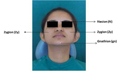
Figure 1: Facial measurement points
Morphological facial height (FH) is the
distance between nasion and gnathion (N-gn). It was measured by spreading
caliper with scale as follows (Figure 2).

Figure 2: Morphological Facial Height.
The fixed tip of the spreading caliper was placed at the
subject’s gnathion and the movable part was moved to place on the nasion. The morphological maximum width of face (FW)
is the distance between the two bilateral zygomatic prominences (zygion to
zygion). It was also measured by spreading caliper with a scale in the
following way (Figure 3).
Figure 3: Morphological Facial Width.
After palpation by fingers, the most lateral point of the zygomatic arch (arcus
zygomaticus) on both sides of the face were located, the ends of spreading
caliper were placed at these points, with enough pressure to feel the bone
under the spreading caliper. The spreading caliper was slightly shifted in the
directions of up and down and back and forth, until the maximum value was
shown.
Facial Index (FI) is the ratio of morphological facial height (FH) and
maximum facial width
(FW) and can be calculated according to the formula: FI = FH/ FW × 100. The values of Facial Index (FI)
were used to determine the incidence of certain facial types according to
Martin-Saller’s scale [3].
The patients were placed in their respective categories
based on their index values. The
subject’s canine and molar relation were recorded with the help of mouth mirror
using the Angle’s classification. Assessment of the antero-posterior
relationship of canine was based on modified Angle’s Classification that
includes three basic classes [4]
Class I: The tip of the maxillary canine lies in the embrasure in between the
mandibular canine and the first premolar.
Class II: The tip of the maxillary
canine lies mesial to the embrasure in between the mandibular canine and
first premolar.
Class III: The tip of the maxillary canine lies distal to the embrasure in
between the mandibular canine and first premolar.
Statistical Analysis
The statistical
analysis was done using SPSS (Statistical Package for Social Sciences) (Version
15) statistical analysis software. The data was subjected to descriptive
analysis for mean, standard deviation, median. A student t-test was applied to
test the significance of two means.
Results
Overall, majority had Euryproscopic type
(53.2%) followed by those having Mesoprosopic type (21.6%), Hypereuryprosopic
type (19%), Leptoprosopic (5.6%) and Hyperleptoprosopic type (0.6%) (Table 1).
However, in males, though majority were Euryprosopic (58.4%) however at next
sequence Hypereuryprosopic and Mesoprosopic types had
equal distribution (18.8% each) followed by Leptoprosopic (3.6%) and
Hyperleptoprosopic type (0.4%) respectively. Among females, although maximum
were Euryprosopic, yet they comprised 46.5% of total females followed by
Mesoprosopic (25.2%) and Hypereuryprosopic (19.2%) types. Leptoprosopic and Hyperleptoprosopic
types comprised the 8.2% and 0.9% of total females enrolled in the study.
Statistically, there was a significant difference between two genders with
respect to facial morphological type (p<0.001).
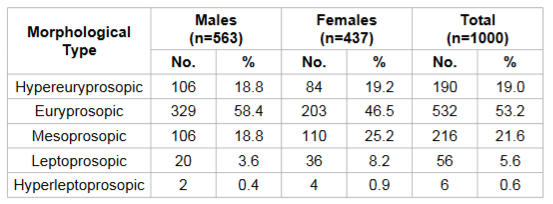
Table 1: Distribution of cases according to Facial Morphological Classification and its association with gender.
Canine relationship for both right and left sides, Class I
was most common. However, for both the sides prevalence of class II and III was
significantly higher in females as compared to males (p<0.05) (Table 2).
Irrespective of facial morphologic type, Class I canine relationship was most
common. Although prevalence of Class I canine relationship was maximum for
Hyperleptoprosopic type as compared to other facial morphologic types yet this
association was not significant statistically for canine relationship of either
side (p>0.05) (Table 3).
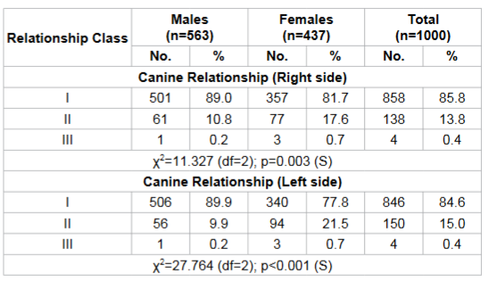
Table 2: Comparison of Canine Relationship between two genders.
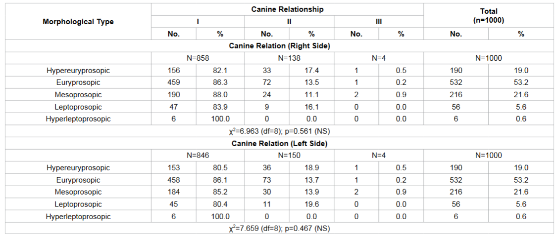
Table 3: Association between facial morphologic types and Canine relationship.
Discussion
Humans are constantly striving to improve their fate. Using facial,
craniofacial, and
maxillofacial surgical techniques, our main aim is to obtain aesthetically
superior results for our patients. To judge the appeal of a face, it is
compared with norms that are today defined by canons or anthropometric
proportions. The availability of values for facial sizes and proportions
enables us to reproduce cosmetically attractive proportions for our patients
[6]. Craniofacial
anthropometry is used for the determination of the morphological
characteristics of the head and face. Face shape is dependent on many factors,
such as gender, race and ethnicity, climate, socioeconomic, nutritional, and
genetic factors. The facial parameters are used to determine the facial trauma,
congenital and traumatic
deformities and easier identification of many congenital malformations. The
collected data can be used in anthropology and forensic medicine for
identification of racial and sexual differences as well as in reconstructive
surgery for facial reconstruction [7].
Diversity and
individuality of people are seen due to variations in the physical shape of
their faces. Studies on craniofacial relations and variations in human will
assist in understanding the frequency and distribution of human morphologies.
Craniofacial anthropometry has become an essential tool for genetic counselors
to identify any dysmorphic
syndromes. Measurements taken from a person can be compared with the normal
values obtained from a reference population, and these deviations from the
normal values can be evaluated [8]. Cephalic and prosopic indices are important
parameters that are used in anthropological studies for showing the variation
between different sex as well as ethnic groups [9].
In this study, the maximum facial height (FH) observed in
males it was 133 mm and in females it was 129 mm. The data were compared
statistically, the difference was found to be significant. Similar result was
obtained from the study done by Jeremic et al. [7] in Central Serbian
population, where they observed facial height to be 121.4 mm in males and 110.8
mm in females. The present study shows that males have higher facial height
than females.
The maximum facial width (FW) in males was 137 mm and in
females it was 135mm. The minimum facial width observed in males was 103 mm and
in females it was 100 mm. On comparing the data statistically, the difference
was found to be significant (p<0.001). These findings were in accordance to
the results obtained by the study done by Young et al. [10], where the maximum facial width was 139.9
mm in bruxers and 131.9 mm in non-bruxers. Jeremic et al. [7] measured facial
widths of 129.1 mm in males and 119.9 mm in females showing that males have
higher facial width than females.
Overall, facial index values ranged from 70.4 to 121% with a
mean value of 89.94 ± 4.54%. In males, the values ranged from 76.6 to 114.6%
with a mean value of 90.16 ± 3.97% whereas in females, the values ranged from
70.4 to 121% with a mean value of 89.65 ± 5.16%. On comparing the data
statistically, the difference was found to be significant (p<0.001).
These findings were
similar to the results obtained by the study of Shetti et al. [11], who
observed mean facial index of 87.19% in males and marginally higher value of
86.71% in females indicating Mesoprosopic facial form.
Similar findings were found in a study which was done by Kurania et al. [12],
the facial index of 89.5% in males and 86.6% in females. In another study it was observed that the
facial index was 85.4% in females and 85.5% in males which is dissimilar to
observations of our study [13]. The probable cause could be that their study
was on a different race (Malaysian Indian).
Overall, as well as for both the genders, majority of
subjects were 18-25 years old. However, proportion of females in age group
18-25 years (83.1%) was higher as compared to corresponding proportion of males
(68.4%) whereas relatively higher proportion of males was aged 26-30 and 31-40
years (20.8% and 10.8% respectively) as compared to females (11.7% and 5.3% respectively). Statistically, there
was a significant difference between two genders with respect distribution in
different age groups (p<0.001). This is in accordance with the study done by
Rexhepi A and Meka V [14] in 2008 on Albanian Kosova population.
In this (present study) study the overall majority had
Euryproscopic facial type (53.2%) followed by those having mesoprosopic type
(21.6%), hypereuryprosopic type (19%), leptoprosopic (5.6%) and
Hyperleptoprosopic type (0.6%).
However, in males, though majority were euryprosopic (58.4%)
however at next sequence Hypereuriprosopic and Mesoprosopic types had equal
distribution (18.8% each) followed by Leptoprosopic (3.6%) and
Hyperleptoprosopic type (0.4%) respectively.
Among females, although maximum were euryprosopic, yet they
comprised 46.5% of total females followed by Mesoprosopic (25.2%) and
Hypereuryprosopic (19.2%) types. Leptoprosopic and Hyperleptoprosopic types
comprised the 8.2% and 0.9% of total females enrolled in the study.
Statistically, there was a significant difference between two genders with
respect to facial morphological type (p<0.001).
Jeremic et al. [7] on
central Serbian population found the dominant facial type Leptoprosopic with an
incidence of 81.7% which was followed by 14.28% of Mesoprosopic and
Hyperleptoprosopic with a frequency of 4% which is different from the result of
our study. In 2003 Golalipour et al. [15] observed the Turkman and Fars
population and found that the dominant and rare facial type was
hypereuriprosopic and leptoprosopic respectively. The findings of this study
were different from the present study in terms of dominant facial type which
was euryprosopic.
In the study done by
Heidari Z et al. [16], they found that in 18-25 years old Baluchi and Sistani
young woman, the dominant and rare facial type was Euryprosopic and
Hyperleptoprosopic respectively in both the populations. This is in accordance
to the present study as in females of 18-25 years old age group, majority of
the subjects were Euryprosopic and Hyperleptoprosopic facial type was found to
be less common.
According to the study done by Rexhepi A and Meka V [14] in
2008, they found that the Leptoprosopic was the dominant facial type
in males followed by Hyperleptoprosopic while in females; Hyperleptoprosopic
was very common in the age group of 18-35 years of Kosova Albanian population.
Hypereuriprosopic was the least common followed by euryprosopic facial type
among both the genders. This result was dissimilar with our study.
The study done by Bayat PD and Ghanbari A [17] in 2009, in Ark, Fars and Turkmen (newborn
population of central Iran and Iranian racial subgroups) found that the
dominant facial type was Hypereuryprosopic for Fars and Ark while Mesoprosopic
for Turkmen. In 2010, Raji et al. [9] found in north-eastern Nigerian population
that the dominant and rarest facial type in both the genders was
Hyperleptoprosopic and Hypereuryprosopic. With respect to the canine
relationship for both right and left sides, Class I was most common. However,
for both the sides prevalence of class II and III was significantly higher in
females as compared to males (p<0.05). When the association between
morphological facial type, canine relationship was observed, it was found that
Class I canine relationship was most common. On comparison of different facial
types, canine relationship, Euryprosopic was found to be dominant facial type
supported by the study done by Young et al. [10]. Irrespective of facial
morphologic type, Class I canine relationship was most common. Although
prevalence of class I canine relationship was maximum for Hyperleptoprosopic
type as compared to other facial morphologic types yet this association was not
significant statistically for canine relationship of either side (p>0.05).
In terms of canine relationship, A significant association between canine relationship
and facial morphologic type was observed at both right and left sides
(p<0.05). On comparing the data, there was no significant association found
between facial morphologic types, canine relationships in both the genders in
different age groups on either side. Only canine relationship association with
facial morphologic type was significant for left side.
Conclusion
The following conclusion may be drawn from the present
study:
• The general facial morphological types
did not show any significant association with canine relationship except for
gender. The age confounded relationship did not show an empirical pattern for
all the age group when evaluated independently.
• Euryprosopic facial type (53.2%) was most common in majority of the subjects
followed by Mesoprosopic (21.6%), Hypereuryprosopic (19%), Leptoprosopic (5.6%)
and the least common was http://edelweisspublications.com/journals/30/
(0.6%).
• Males and females both showed the majority of 58.4% and 46.5% respectively of
Euryprosopic facial type on comparing the data with facial type to gender. This
showed the significant difference between two genders with respect to the
facial morphology.
• The canine relationship showed Class I in both the genders while Class II and
Class III were slightly increase in females.
• The association between morphological facial type, molar and canine
relationship was observed and found that the class I molar and canine
relationship was most common.
• In males, the prevalence of Class I canine relationship
was significantly higher in Mesoprosopic and Hyperleptoprosopic as compared to
other types for both the sides.
• In females, there were no significant association found between morphological
facial types, canine relationship on either side.
• There was no significant association between facial morphologic types and
canine in both the genders at either side. The canine relationship association
with facial morphologic type was significant for left side.
References
1. Kumar M, Lone MM.
The study of facial index among Haryanvi adults. (2013) IJSR 2:51-53.
2. Hegde C, Lobo NJ, Prasad KD. A cephalometric study to
ascertain the use of nasion as a guide in locating the position of orbitale as
an anterior reference point among a population of South Coastal Karnataka. (2013)
Contemporary Clinical Dentistry 4: 325-330.
3. Al-Yessary AS, Al-Fatlawi FA. Facial profile, occlusal
features and treatment need for a sample of Karbalaa governorate students aged
14 years. (2011) J Bagh College Dentistry 23.
4. AL-Maqtari RA, Kadir RA, Awang H, Zam Zam NM. Assessment
of malocclusion among yemeni adolescents using canine and incisor
classifications. (2012) J Advan Med Res 2:153-159.
5. Behbehani F, Roy R, Al-Jame B. Prevalence of asymmetric
molar and canine relationship. (2012) Eur J Orthod 34: 686-692.
6. Vegter F. Clinical anthropometry and canons of the face
in historical perspective. (2000) Plast Reconstr Surg 106:1090-1096.
7. Jeremic D. Anthropmetric study of the facial index in the
population of central Serbia. (203) Arch Biol Sciences 5: 1163-1168.
8. Mostafa A, Banu LA, Rehman F, Paul S. Craniofacial
anthropometric profile of adult bangladeshi buddhist chakma females. (2013)
Journal of Anthropology 1-7.
9. Raji JM, Garba SH,
Numan AI, Waziri MA, Maina MB. Morphological evaluation of head and face shapes
in a north - eastern nigerian population. (2010) Aust J Basic and Appl Sci 4:
3338-3341.
10. Young DV, Rinchuse DJ, Pierce CJ, Zullo T. The
craniofacial morphology of bruxers versus non-bruxers. (1994) Angle Orthod 69:
14-18.
11. Shetti VR, Pai SR, Sneha GK, Gupta C, Chethan P, et al.
Study of prosopic (facial) index of indian and malaysian students. (2011) Int J
Morphol 29:1018-1021.
12. Kurnia C, Susiana, Husin W. Facial indices in chinese
ethnic students aged 20 22. (2012) Journal of Dentistry Indonesia 19: 1 4.
13. Ngeow WC, Aljunid ST. Craniofacial anthropometric norms
of malaysian indians. (2009) Indian J Dent Res 20: 313-319.
14. Rexhepi A, Meka V. Cephalofacial morphological
characteristics of albanian kosova population. (2008) Int J Morphol 26:
935-940.
15. Golalipour MJ, Haidari K, Jahanshahi M, Farahani RM. The
shapes of head and face in normal male newborns in south-east of caspian sea
(irangorgan). (2003) J Anat Soc India 52: 28-31.
16. Heidari Z, Sagheb HRM, Mugahi MHN. Morphological
evaluation of head and face in 18-25 years old women in southeast of iran.
(2006) J Med Sci 6: 400-404.
17. Bayat PD, Ghanbari A. The evaluation of craniofacial
dimensions in female arak new borns (central Iran) in comparison with other Iranian
racial subgroups. (2009) Eur J Anat 13: 77-82.


