Introduction
Leiomyosarcomas of the penis are very rare and less
than 50 cases of the disease have been reported in the literature. In view of
this majority of clinicians would not have come across a case of the disease
and perhaps may not have a high index of suspicion of the disease. Despite the
rarity of leiomyosarcoma
of the penis, it is the second commonest sarcoma of the penis. Leiomyosarcoma
of the penis has been classified into superficial and deep sub-types depending
upon the site of origin of the tumor [1,2].
Leiomyosarcomas of the penis could arise from
· The
dartos muscle of the prepuce and shaft of the penis.
· The
erector pilorum muscle of the shaft of the penis.
· The
muscle wall of the superficial vessels.
· The
smooth muscle of the deep vessels which constitute the corpora cavernosa and
the corpus spongiosum.
Primary
leiomyosarcoma of the penis may mimic other lesions of the penis including
Kaposi sarcoma, Leiomyoma, melanoma with storiform growth, myointimoma and sarcomatoid carcinoma
of the penis and hence a careful histopathology and immunohistochemistry
staining studies of penile lesions are required to confirm the diagnosis of
primary leiomyosarcoma of the penis [3].
The ensuing review and update of the literature on
primary leiomyosarcoma of the penis is divided into two parts including (A)
Overview and (B) Miscellaneous narrations and discussions from some case
reports, case series, and some studies related to primary leiomyosarcoma of the
penis.
Aim
To review and update the literature on primary
leiomyosarcoma of the penis.
Method
Internet data bases were used including: Google,
Google Scholar, Yahoo, and PUBMED. The search words that were used included
Leiomyosarcoma of the penis, penile leiomyosarcoma,
Fifteen references were identified which were used to write the review and
update of the literature.
Results
Overview
Definition/General
Statements: Leiomyosarcoma of the Penis (LOP) is a malignant
smooth muscle tumor of the penis like smooth muscle malignant tumors elsewhere
in the body [3]. LOP can arise from:
· The
dartos muscle of the prepuce and shaft of the penis.
· The erector pilorum muscle
of the shaft of the penis.
· The
muscle wall of superficial vessels.
· The
smooth muscle of the deep vessels which constitute the corpora cavernosa and
corpus spongiosum.
Epidemiology:
PLOP
is an extremely rare malignancy that affects the penis and less than 50 cases
of primary leiomyosarcoma of the penis have been reported in the literature
therefore majority of clinicians globally would not have encountered a case of
the disease [3]. LOP is the second commonest sarcoma of the penis after Kaposis sarcoma of the
penis [3]. It has been documented that the ages of patients who have been
reported to have had PLOP have ranged between 43 years and 62 years and the
mean age of patients at the time of the initial diagnosis of PLOP was 51 years [3].
Sites:
It
has been iterated that PLOP usually tends to affect the shaft of the penis but
PLOP can afflict any site of the penis [3].
Presentation:
PLOP
may present with a history of variable duration of:
· Lump
or mass anywhere on the penis including the prepuce, glans of penis, and
anywhere along the shaft of the penis.
· Ulcer
anywhere on the penis (prepuce, glans, shaft of penis).
· Nodule(s)
on the penis.
· Pruritus
on penis.
· Dysuria
on rare occasions when a deep LOP affects the urethra.
· Voiding
difficulties on rare occasions when a deep LOP has invaded or compressed the urethra.
· Induration
of the penis.
Clinical
Examination Findings:
· The
general and systematic examination of most patients would tend to be normal
unless they have other medical problems not related to LOP.
· Lump
anywhere on the penis.
· Ulcer
usually with raised edges anywhere on the penis.
· Nodule
or nodules on the penis.
· In
majority of cases there will be no palpable inguinal lymph node enlargement.
· Inguinal
lymph node
enlargement may be observed on rare occasions if the tumor is in an advanced
stage.
Laboratory
Investigations
Urine:
Urinalysis,
urine microscopy and urine culture and sensitivity are general tests that are
undertaken in the general assessment of patients who have LOP but the results
on the whole would tend to be normal; nevertheless, if there is any evidence of
urinary tract infection it would be treated according to the antibiotic
sensitivity pattern in order to improve the general condition of the patient
prior to treating the LOP.
Bacteriology
culture of discharging penile ulcer as may be required: On
rare occasions an LOP may present as a penile ulcer and if there is any
discharge from the ulcer and an infection is suspected swabs of the discharge
are taken for microscopy
and culture and if there is any evidence of infection it would be treated accordingly
to improve the general condition of the patient.
Hematology
Blood Tests: Full blood count and coagulation screen are routine
tests that are undertaken in the general assessment of patients who have LOP
and generally the results would tend to be normal but if there is any
abnormality it would be investigated accordingly and the most appropriate
treatment would be given to improve upon the general condition of the patient.
Biochemistry
Blood Test: Serum urea, creatinine,
electrolytes, blood glucose, and liver function tests are routine tests that
are undertaken in the general assessment of patients who have LOP and generally
the results would tend to be normal but if there is any abnormality it would be
investigated accordingly and the most appropriate treatment would be given to
improve upon the general condition of the patient.
Radiology
Investigations
Chest
X-ray:
Chest X-ray is a
routine test that tends to be undertaken in the routine assessment of patients
who have LOP and this can be combined with ultrasound scan of the abdomen and
pelvis as well as the penis in the follow-up assessment of patients who have
undergone treatment for LOP to ascertain if they have developed metastases but
this has been superseded in the developed countries by utilization of computed
tomography scan/magnetic resonance imaging scan of thorax, abdomen and pelvis.
In parts of the world where CT and MRI scan facilities are not available,
chest-ray and ultrasound scans are routinely undertaken in the follow-up
assessment of patients.
Ultrasound
Scan: Ultrasound
scan of penis, abdomen and pelvis tends to be undertaken as part of the initial
assessment of patients who have LOP and this can also be undertaken in the
follow-up assessment of patients to determine whether or not local recurrence,
lymph node enlargement or intra-abdominal and pelvis metastases have developed.
Most often in the developed countries staging of the tumor and follow-up
assessments are undertaken by utilization of CT/MRI scan of thorax, abdomen,
and pelvis.
Computed
Tomography (CT) Scan: CT scan of thorax,
abdomen and pelvis including the penis tends are to be undertaken as part of
the full initial staging of patients who have PLOP. CT scan of thorax, abdomen
and pelvis also forms part of the thorough follow-up protocol in the assessment
of patients who have undergone treatment for PLOP.
Magnetic
Resonance Imaging (MRI) Scan: MRI scan of thorax, abdomen and
pelvis including the penis tends to be undertaken as part of the full initial
staging of patients who have PLOP.MRI scan of thorax,
abdomen and pelvis also forms part of the thorough follow-up protocol in the
assessment of patients who have undergone treatment for PLOP.
Positron
Emission Tomography (PET)/CT Scan: PET/CT scan is an
investigation that can be undertaken in the follow-up assessment of patients
who have undergone treatment for PLOP to investigate whether there is any
metastatic or recurrent lesion anywhere in the body.
Isotope
Bone Scan: Isotope bone scan
tends to be undertaken to ascertain if bone metastasis has developed in
patients who have undergone treatment for LOP.
Clinical
Features
· It has
been stated that with regard to PLOP the tumor would more likely tend to be a
lesion that is superficial (superficial to or above the tunica albuginea) than
deep seated [3].
· It has
also been iterated that usually pursuant to excision of PLOP, the PLOP lesion
quite often tends to recur locally.
· It has
been stated that with regard to symptoms, superficial PLOPs tend to be
asymptomatic but on the other hand deep seated PLOPs tend to manifest with
dysuria or difficulty with micturition [3].
Diagnosis:
The
diagnosis of LOP can be established through pathology examination of specimen
of the penile lesion in various scenarios as follows:
· Wedge
biopsy of the penile lesion.
· Aspiration
of the lesion for cytopathology examination.
· Examination
of the fully excised lesion of the penis.
· Examination
of the partial amputation specimen.
· Examination
of the total penectomy
specimen.
Prognostic
Factors: It
has been iterated that favorable factors associated with the outcome of PLOP
pursuant to treatment include [1,3].
· When
the depth of the tumor is less than or equal to 2 cm.
· When
the size of the tumor is less than or equal to 5 cm.
Treatment
· With
regard to treatment of PLOP, it has been stated that wide local excision
ensuring complete excision of the tumor with tumor-free and clear surgical
excision margin would be sufficient treatment to ensure all deep lesions are
removed [1,3].
· Wide
surgical excision of the tumor would tend to be feasible with regard to superficial lesions [3].
· Some
cases of extensive deep seated LOPs that are large and invading the urethra or
corpora may necessitate partial amputation of the penis or perhaps total
penectomy depending upon the site of the penis involved to ensure complete
excision of the tumor.
· It has
been stated that circumcision could prove to be sufficient treatment for cases
of leiomyosarcoma that is only localized to the foreskin (prepuce) [4].
· It has
been stated that utilization of adjuvant chemotherapy and radiotherapy do not
have any role to play generally in the management of LOP. However, in view of
the fact that the deep-seated type of LOP tends to be associated with the
development of tumor recurrence as well as distant metastases, utilization of a
combination of local radiotherapy to the pelvis and systemic chemotherapy could
be effective as adjuvant treatment [5].
Macroscopic
Examination Features
· It has
been stated that gross examination in cases of PLOP does show tumor that has
measured between 0.5 cm and 6.0 cm and that the median size has been 1.5 cm [1,3].
· It has
been documented that PLOPs that have been reported have most often been
superficial tumors [1,3].
· It has
been iterated that gross examination of PLOP does reveal tumors that are
white-tan-grey, and firm as well as the tumors tends to have irregular borders.
[1,3].
Microscopic
Examination Features: The microscopic examination features of
PLOP have been summated as follows [1,3]:
· Microscopic
examination of the tumor does show interlacing fascicles of spindled-cells that
have abundant eosinophilic cytoplasm as well as focal juxta-nuclear vacuoles
and blunt ended nuclei.
· Microscopic
examination of PLOP also does show a degree of atypia which tends to range
between moderate atypia to severe atypia.
· Microscopic
examination of PLOP on rare occasions does show atypical multi-nucleated giant
cells, and focal necrosis.
· Mitoses
tend to be found easily upon microscopic examination of PLOP and the mitoses
could be many.
Immunohistochemistry
Studies
Positive
Staining:
It has been iterated that immunohistochemistry staining studies in cases of
PLOP tend to show that the tumors do exhibit positive staining for the ensuing tumor markers [1,3]:
· Desmin
(positive staining).
· Muscle
specific actin (positive staining).
· Alpha
smooth muscle actin (positive staining).
The tumor cells would also tend to exhibit positive
staining for vimentin.
Negative
Staining:
The tumor cells would also tend to exhibit negative staining for keratin.
Electron
Microscopic Examination: It has been stated that electron
microscopy examination of leiomyosarcoma of the penis would tend to show the
following: Myofibrils, dense bodies, and abundant pinocytic vesicles
tend to be noted, and a continuous basal lamina tends to be present around
majority of the tumor cells [6].
Differential
Diagnoses: Some
of the stipulated differential diagnoses of PLOP have been summated as follows
[3]:
Kaposi
Sarcoma (Nodular): These tumors tend to be located
superficially and microscopic examination of the Kaposi Sarcoma tumors tend to
show slit-like spaces associated with many erythrocytes, no evidence of atypia.
Immunohistochemistry staining studies of Kaposi sarcoma of the penis tends to
show positive staining for:
· HHV8
· CD31
· CD34
· Factor
VIII
Leiomyoma: Leiomyomas
of the penis are extremely rare and the microscopic examination features of
these benign lesions tend to be similar to the features of leiomyomas elsewhere
within the body. Microscopic examination of leiomyoma of the penis does show
rare or no mitotic figures, no atypia and no evidence of tumors necrosis.
Melanoma
with Storiform Growth: Melanoma with storiform growth within
the penis does look like typical melanoma elsewhere within the body.
Immunohistochemistry staining studies of melanoma with storiform growth pattern
does show positive staining for melanocyte markers including:
· HMB45
· Melan
A
Myointimoma: Myointimona
tends to be associated with more plexiform growth pattern and microscopy
examination of myointimoma does show no evidence of nuclear atypia and
immunohistochemistry staining studies show minimal reactivity for desmin.
Sarcomatoid
Carcinoma:
sarcomatoid carcinoma of the penis does consist of squamous epithelial nests
within the tumour mass and associated areas of PeIN. Immunohistochemistry
staining studies in sarcomatoid carcinoma of the penis does exhibit positive
staining for:
· Pankeratin
· Keratin
34beta E12
· p63
In sarcomatoid carcinoma of the penis
immunohistochemistry staining studies show negative staining for muscle
markers.
Outcome
· Superficial
PLOPs tend not to recur after complete wide excision of the tumors ensuring no
residual tumor but some very large superficial PLOPs that are associated with a
high mitotic activity do recur and would require careful follow-up. Presence of
tumor close to the excision margin may subsequently result in the subsequent
development of recurrence.
· Some deep
PLOPs despite adequate radical surgery alone or plus chemotherapy /radiotherapy
do tend to be associated with the subsequent development of local recurrence,
metastasis and death of the patient.
· There
is need to investigate the use of immunotherapy and new chemotherapy
medicaments to ascertain if this would lead to improvement in the outcome of
patients and the disease.
Miscellaneous
Narrations of Some Case Reports, Case Series and Studies Related to
Leiomyosarcoma of the Penis
Fetsch, et al. in 2004 stated that PLOP was rare and
by then less than 30 cases of PLOP had been reported in the English literature [1].
Fetsch, et al. described the clinical, histopathological, and immunohistochemistry studies
findings of 14 cases of PLOP they had retrieved [1]. The mean age of the
patients was 51 years and the ages of the patients had ranged between 43 years
and 62 years at the time of the initial resection of the tumors. With regard to
the location of the tumor, they reported the ensuing: The tumor had involved
the prepuce in 1 case, the prepuce as well as the distal shaft in 1 case, the
circumcision scar line in 2 cases, the circumcision scar line and distal shaft
in 1 case, the shaft of the penis in 5 cases, the base of the penis in 3 cases,
and the penis the site of which was not otherwise specified in 1 case [1]. The
median size of the lesions was 1.5 cm and the sizes of the lesions had ranged
between 0.5 cm and 6.0 cm in their maximum dimension.
Nine of the tumors were in a superficial location of
the penis, the position of tumors were stated as indeterminate in 2 cases, and
3 cases were stated to be in the deep part of the penis. With regard to
symptoms, the superficial tumors were reported to be relatively asymptomatic
and seven of the cases had been reportedly present for 1 year to longer than 20
years with a median duration of 5 years before medical attention was sought. In
comparison, one deep-seated penile lesion did cause dysuria and difficulty in micturition,
which had prompted the patient to look for a clinical medical opinion with only
a few months of the apparent onset of the symptoms. With regard to
histopathology examination features of the tumor, all of the tumors were found
to have contained smooth muscle cells that had both cytological examination
features of atypia as well as mitotic activity.
There were available the results of
immunohistochemistry staining studies for nine of the tumors and it was
documented that the immunohistochemistry studies had shown that all of the tumors
had exhibited positive immunohistochemistry staining for desmin.
With regard to treatment, all of the patients had in
the first instance undergone a local procedure. With regard to follow-up, there
was data available for 9 out of the 14 patients which amounted to 64% of the
patients and the median follow-up for the patients was documented as 12 years
and 11 months. Three patients did develop multiple local recurrences of between
2 and 4 recurrences. 2 of the aforementioned patients had ultimately undergone
a wide local excision or partial penectomy, and both patients were alive and
well at their last follow-up. On the contrary, one patient, who had developed 4
local penile recurrences and who had refused a penectomy, did develop a distant
metastasis 10 months pursuant to the development of the 4th recurrence.
Fetsch, et al. made the ensuing iterations [1]:
· The
best predictors of outcome of PLOP include the depth of the tumor, and the size
of the tumor.
· Superficial
PLOPs are best managed by means of wide local excision of the tumor whenever
local excision is technically possible.
· Tumors
that have a deep-seated component could require more aggressive interventional
procedure treatments that would ensure complete removal of the tumors.
Sundersingh, et al. reported a 56-year-old man who
had presented with a one and half months history of pain and swelling of his
penis [2]. His clinical examination revealed a 2 cm x 2 cm indurated ulcer that
had raised edges and a hard plaque-like thickening which had involved the
distal shaft of his penis upon the right side and dorsal aspect of his penis that
had extended onto his glans penis. No inguinal or iliac lymph nodes were
palpable during his clinical
examination.
He had ultrasound scan of his abdomen and pelvis
which were normal. He also had CT scan of his thorax which was normal. He had
wedge biopsy of the lesion which did show a tumor which had consisted of
fascicles of spindled-cells that had eosinophilic cytoplasm
and hyper chromatic elongated cigar-shaped nuclei that had blunt ends which had
exhibited moderate to marked atypia of nuclei. There were 0 to 2 mitoses per
high-power field (Figure 1).
Immunohistochemistry staining studies of the tumor
did show that the tumor cells had exhibited positive staining for: vimentin,
muscle actin, desmin, and Smooth Muscle Actin (SMA) (see Figure 2). The immunohistochemistry
staining studies also showed that the tumor cells had exhibited negative
staining for keratin. The proliferative activity of the tumor which was
ascertained by Ki-67 index was 40%. Based upon the histopathology and
immunohistochemistry staining features of the tumor a diagnosis of grade 3
leiomyosarcoma of the penis was made. He did undergo total penectomy.
Macroscopic examination of the specimen demonstrated an ulcerated tumor that
measured 2 cm x 2 cm which had involved the dorsal area of the right distal
half of his penis and which had extended into his glans penis. Examination of
the cut surface of the tumor showed a grey-white, firm, deep-seated tumor which
had measured 3.5 cm x 3 cm x 3 cm.
Histopathology examination of the specimen revealed
features that was suggestive of LOP grade 3 which had involved the corpora
cavernosa as well as the corpus spongiosum (see Figure 3). The urethral mucosa did not contain any tumor and areas
of necrosis as well as degeneration were visualized. The surgical resection
margins were free of tumors. Sundersingh, et al. made the following iterations:
[2]
· Leiomyosarcoma
of the penis is the second commonest sarcoma of the penis [2].
· Leiomyosarcoma
of the penis has been classified into (a) superficial and (b) deep sub-types of
leiomyosarcoma depending upon the site of origin of the tumor [1].
· Leiomyosarcomas
of the penis can arise from (a) the dartos muscle of the prepuce and shaft of
the penis, (b) the erector pilorum muscle of the shaft of the penis, (c) the
muscle wall of superficial vessels, and (d) the smooth muscle of the deep
vessels which constitute the corpora cavernosa and corpus spongiosum.
Leiomyosarcoma tumors that arise from the former three sites including (a), (b)
and (c) do constitute the superficial types of leiomyosarcoma of the penis, and
the last site (d) constitute the deep leiomyosarcomas of the penis.
· Deep
seated LOP do manifest as large, rapidly growing, poorly circumscribed, firm
masses that have the tendency to invade the urethra [7].
· It has
been iterated that surgery does remain the mainstay of treatment and that small
deep-seated tumors that are located within the distal shaft or glans of the
penis could be effectively treated by means of partial penectomy, whereas,
large deep-seated tumors, especially those tumors that are situated at the root
of the penis would require treatment by means of total penectomy (total
amputation of the penis) [1].
· It has
been stipulated that the undertaking of regional lymph node dissection would
usually not be indicated in view of the fact that nodal metastases tend not to
be common [4].
· It has
been iterated that utilization of adjuvant chemotherapy and radiotherapy do not
have any role to play generally in the management of LOP. Nevertheless, in view
of the fact that the deep-seated type of LOP tends to be associated with the
development of tumor recurrence as well as distant metastases, utilization of a
combination of local radiotherapy to the pelvis and systemic chemotherapy could
be effective as adjuvant treatment [5].
· The
depth of the tumor as well as the size of the tumor would appear to be the best
predictors of clinical prognosis and deep-seated large tumors often tend to be
associated with poor outcome [1].
· Histopathology
prognostic factors of primary leiomyosarcoma (PLOP) do include (a) tumor growth
pattern depending upon if the tumor is circumscribed or infiltrative, and (b)
high mitotic count of greater to 10 mitoses per/10 high-power fields tends to
be associated with inferior outcome, (c) grade 3 tumors tend to have inferior
prognosis [1].
· Their
patient was alive six months pursuant to his surgery.
Dominici, et al. reported a 53-year-old man who
underwent postectomy for a firm nodule in his prepuce [7]. He did experience
four years subsequently local recurrence of tumor which was successfully
treated by means of partial
penectomy. Katsikas, et al. reported a 78-year-old man.
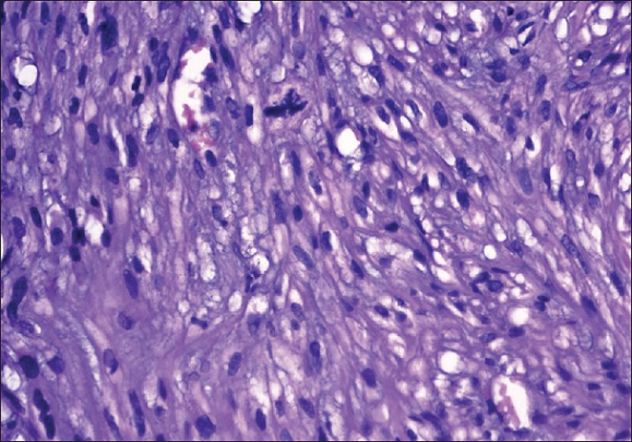
Figure 1:
Fascicles of hyper chromatic spindle cells exhibiting cigar-shaped nuclei with
an atypical mitosis (H and E x 40) [2].
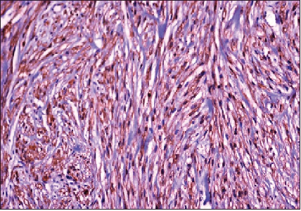
Figure 2: Tumor
cells are positive for smooth muscle actin. (IH x 40) [2].
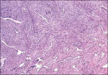
Figure 3: Tumor
involving corpora cavernosa (H and E x 5) [2].
who had presented with a 3-year history of gradual
painless swelling of his penis [8]. His clinical examination revealed a
non-mobile hard mass which had involved the base and mid-shaft of his penis.
His glans penis was normal and there was no obvious palpable regional lymph
node enlargement. The results of his routine hematology and biochemistry blood
tests were normal. He had a CT scan which showed a soft tissue mass lesion which
had involved the corpora cavernosa of his penis and no evidence of metastasis. He
underwent radical penectomy and perineal urethrostomy
under the provisional diagnosis of sarcoma of the penis. Gross examination of
the specimen showed a tumor which had measured 8 cm x 8 cm x 14 cm which had
arisen from the corpora cavernosa and the nature of the tumor had been such
that the corpora could not be differentiated from one another. The urethra and
glans penis were free of tumor (see Figure
4).
Microscopic examination of the tumor revealed
features of a high grade sarcoma which had consisted of neoplastic
spindled-shaped cells that had eosinophilic cytoplasm that had been arranged in
fascicles. The cells were found to have nuclear atypia and hyper chromatic nuclei
as well as the mitotic rate of the tumor was five mitoses per high-power field.
Immunohistochemistry staining studies of the tumor showed that the tumor cells
had exhibited positive staining for vimentin and SMA (see Figure 5). The tumor cells also exhibited negative staining for
desmin and S-100 protein.
At his 2-year post-operative follow-up, he was well
with no evidence of local recurrence or distant metastasis. Katsikas, et al.
stated the following [8]:
Note:
The urethra is free of invasion; the tumor size is estimated to be
approximately 18 cm.
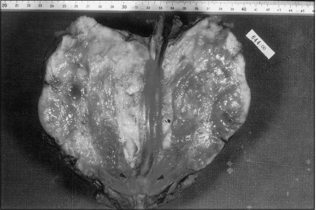
Figure 4:
Macroscopic view of the excised tumor, arrowheads indicating the glans penis at
the bottom [8].
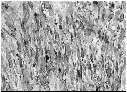
Figure 5: On
immune histochemical stains, strong positivity for Smooth Muscle Antigen (SMA,
100) can be seen [8].
· Pratt
and Ross were the first authors who had classified leiomyosarcoma of the penis
into two distinct clinical and pathological entities of superficial and deep
leiomyosarcomas [9].
· The
options of treatment for leiomyosarcoma of the penis include: (a) Surgery, in
the form of local excision, amputation whether partial or total or radical
penectomy, (b) radiotherapy, or (c) chemotherapy.
· Surgery
should be aimed at the excision of the tumor mass. It has been stated that
amputation would tend to be the most effective surgical treatment to prevent
recurrences for both superficial and deep leiomyosarcomas of the penis;
nevertheless, the approach would tend to be individualized, and in view of the
fact that superficial tumors would tend to manifest in younger men, these cases
could be managed by means of local excision of the tumor with tumor negative
surgical margins whenever it is possible [4].
· Deep
lesions tend to be most appropriately treated via a more aggressive approach by
means of amputation
for distal lesions, or radical penectomy for the middle or proximal penile
shaft of penis lesions.
· It has
been iterated that pre-operative or post-operative external beam radiotherapy
had not been proven to be of value with regard to its value in the treatment of
leiomyosarcomas of the penis or in increasing the survival rates of patients
[10].
· At the
time of the report of their case, chemotherapy with anthracyclines or etoposide
had provided poor results and unfortunately, ongoing trials at the time of
publication of their article with utilization of newer taxanes had not been
successful with regard to the treatment of leiomyosarcomas of the uterus [11].
· However,
both radiotherapy and chemotherapy could be utilized for palliation treatment
with regard to leiomyosarcoma of penis recurrences which would not be amenable
to surgical treatment.
· The
effectiveness of radiotherapy and chemotherapy is debatable and the lack of
large series of cases of leiomyosarcoma of the penis would make conclusions
related radiotherapy and chemotherapy insecure.
· In
view of the small number of cases of PLOP so far, the best approach to the
management of these PLOPs would require collaboration between the urologist,
the pathologist, the radiotherapist, and the medical oncologist in order to
provide optimum results in the best interest of the patient.
Some people could argue that the case was reported
with a short period of follow-up and therefore the long-term outcome of the
patient would not be known. They could further argue that considering that
deep-seated leiomyosarcomas of the penis potentially could be associated with the
subsequent long-term development of metastasis they would have recommended from
the hind sight utilization of adjuvant radiotherapy and chemotherapy as
prophylactic treatment for the prevention of future recurrence. Other
clinicians would support the sole treatment of radical penectomy alone plus
perineal urethrostomy only.
The reasons for supporting radical surgery alone
include the following: (a) There were no enlarged regional lymph nodes; (b) the
patient had a long 3-years history of a penile mass which would indicate a
natural slow-growing tumor. (c) The mitotic rate was 5 mitoses per high-power
field which would indicate a slow-growing malignancy with little
chance of exhibiting an aggressive biological behavior and if the mitotic rate
was higher that would indicate a potential of the tumor to be an aggressive tumor
and for these reasons it was good practice to avoid adjuvant therapy which
could also be associated with side effects. Nevertheless, the factor that could
allude to possible potential for the subsequent development of late metastasis
or recurrence is the large size of the tumor.
Nanri, et al. reported a 27-year-old man who had
presented with a mass at the root of his penis [5]. He underwent biopsy of the
lesion and pathology examination of the specimen showed features consistent
with the diagnosis of leiomyosarcoma of the penis. The tumor was categorized
clinically and pathologically as deep-seated leiomyosarcoma. He had retrograde
urethrogram which showed compression of the anterior urethra but no invasion of
the urethra.
He had MRI scan which showed a solid mass which had
invaded the corpus cavernosum and the corpus spongiosum. No distant or lymph
node metastases were demonstrated. He underwent total penectomy and inguinal
lymph adenectomy. During the procedure, there was evidence of intravenous tumor
extension into the deep dorsal vein of the penis. Pathology examination of the tumor
showed spindled-shaped cells that were arranged in interlacing fascicles, which
did confirm the diagnosis of leiomyosarcoma of the penis. No evidence of lymph
node involvement by the tumor was found on pathology examination of the
specimen.
He received two cycles of combination chemotherapy
which had consisted of mesna, doxorubicin,
ifosfamide and dacarbazine (MAID regimen). He developed a local recurrence 3 months
following his surgery and following this the recurrent tumor had quickly extended
to involve the prostate gland, the seminal vesicle, the urinary bladder, and
the rectum. The tumor subsequently metastasized to his lungs and bone. He died
14 months pursuant to his surgery. Nanri, et al. stated that their case was the
45th case of leiomyosarcoma of the penis to be reported in the
literature [5]. Lessons that would need to be learnt from this case report is
that deep-seated leiomyosarcomas of the penis that invade the dorsal vein of
the penis are aggressive tumors which could quickly metastasize and the patient
was treated aggressively but there was no response to the combination
chemotherapy.
This experience would indicate that there is need
for Urologists, oncologists, pharmacotherapy research workers, and pathologists
to undertake research work that would identify new chemotherapy medicaments
that will effectively destroy microscopic metastatic leiomyosarcoma cells to
prevent them developing into overt metastases. Additionally, some people could
argue that considering that primary deep-seated leiomyosarcoma which has
invaded the dorsal vein of the penis is an aggressive tumor it should also be
treated with adjuvant radiotherapy with the aim of destroying microscopic tumor
cells whether this would be effective or not cannot be ascertained and the
answer would be known if such patients are entered into a global multi-center
trial. Furthermore, it would be argued that patients who have deep-seated
primary leiomyosarcoma with invasion of the deep dorsal vein of the penis
should be entered into a multi-center global trial of immunotherapy to
ascertain if immunotherapy would prevent the development of metastasis.
Gonzalez, et al. reported a 39 year old man who had
had frenuloplasty
in 1990 who was a smoker and who had presented in December 2009 with an
enlargement of a tumor on the ventral aspect of his penis of 1 year duration
which was associated with pruritus [12]. His clinical examination showed a
palpable tumor which had measured 1 cm within the frenulum of his prepuce,
adjacent to his external urethral meatus.
The tumor mass was mobile, and painless. There was
no evidence of inguinal lymph node enlargement. The lesion was excised under
local anesthesia. Pathology examination of the tumor revealed features that
were consistent with the diagnosis of leiomyosarcoma in that it showed long
spindled-shaped tumor cells and that the proliferating cells exhibited nuclear
atypia and mitotic rate of 2 per 10 high-power fields. The Ac. Ki-67 proliferation
index was 25% (see Figures 6 and 7).
He had staging CT scan of thorax, abdomen and pelvis which was normal. He was
well at his 3 years and six months follow-up with no evidence of local
recurrence or distant metastasis.
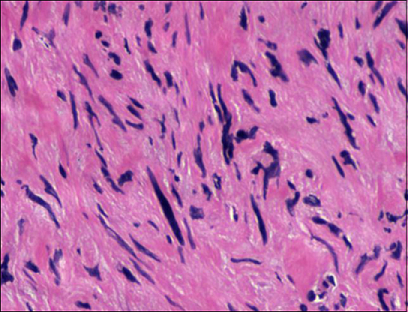
Figure 6:
Spindle cells with clear nuclear pleomorphism and high mitotic index [12].
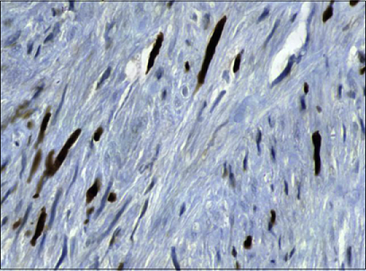
Figure 7:
Immunohistochemistry Ac. Ki767 demonstrating the high proliferating index [12].
It would be argued that the treatment of local
excision of the tumor with wide margin ensuring there is no residual tumor is
adequate treatment for the superficial primary leiomyosarcoma of the penis.
Even though some Superficial
Primary Leiomyosarcomas of the Penis (SPLOPs) could recur locally
subsequently, the factors that are in favor of the adequacy of local excision
of the tumor in the reported case is the small size of the tumor, the superficial
nature of the tumor and the low mitotic rate of the tumor of 2 per 10 high-power
fields which would indicate a possibly non-aggressive tumor. A lesson learnt
from this case report is that despite the patient noticing a lump in his penis
associated with pruritus, he only reported the case one year from the onset of
his symptoms.
Based upon this experience it would be important
that efforts are made globally to educate males about the possibility of
malignancies developing on the penis and all males who notice a lump or an
ulcer on their penis should seek early medical assessment. In some parts of the
developing world, there are very few pathologists and some hospitals do not
have pathologists and for that reason all surgical specimens tend to be sent to
the Regional or National hospitals for pathology examinations. Clinicians need
to be advised to send all excised surgical specimens for pathology examination
to be absolutely sure about the nature of the lesions they excise and no
excised lesion should be thrown away. DCruze, et al. reported a 59-year-old man
who had presented with an ulcer-proliferative growth at the tip of his penis
which he had noticed over the preceding one month [13].
The lesion was documented to be progressive and
associated with pain. He did not have any history of bleeding, weight loss, or
loss of appetite. His general and systematic examinations were normal.
Examination of his genitalia revealed a 4 cm x 4 cm ulcer-proliferative growth
on the tip of his penis. There was no evidence of lymph node enlargement. The
results of his routine hematology and biochemistry blood tests were within
normal range. He had a chest radiograph which was normal. He had ultrasound
scan of his abdomen and pelvis and renal tract which were normal. He had wedge
biopsy of his penile lesion and pathology examination of the specimen showed
features of a high-grade fascicular spindled-cell sarcoma associated with a
mitotic rate of 32 per 10 high-power fields.
Immunohistochemistry staining studies of the tumor
showed that the tumor cells had exhibited strongly positive staining for
vimentin and SMA as well as focal positive staining for S-100. The tumor cells
upon immunohistochemistry staining had exhibited negative staining for CK and
CD34. Based upon the histopathology and the immunohistochemistry staining
features of the tumor, a diagnosis of a high-grade leiomyosarcoma of the penis
was made. He did undergo partial penectomy. The corpus spongiosum was not
involved by the tumor. At his 11 month follow-up he presented with a focal
non-healing wound at the site of his previous operation that was biopsied. Histopathology
examination of the specimen showed inflammatory granulation tissue with no evidence
of recurrent tumor. Since the case was reported with 11 months follow-up one
cannot know the long-term outcome of the disease. DCruze, et al. stated that
only 46 cases of primary leiomyosarcoma of the penis had been reported in the
English literature prior to the report of their case [13]. Khobragade, et al. reported
2 cases of primary leiomyosarcomas of the penis as follows [14]:
Case 1: A
26-years-old man had manifested with progressively increasing painless swelling
at the base of his penis of 3 months duration. His clinical examination
revealed a 3 cm x 3 cm, firm swelling at his penoscrotal junction
that was free from the underlying pubic bone and no evidence of inguinal lymph
node enlargement. He had MRI scan which demonstrated a 4.7 cm x 3.7 cm x 5.4 cm
soft tissues mass that was lobulated and which had involved the left corpus
cavernosum with no evidence of lymphadenopathy. Fine needle aspiration cytology
examination of the specimen was undertaken which had suggested a spindled-cell tumor.
Through an inguinoscrotal incision, the mass was excised with the excision of
the left corpus cavernosum. In view of the fact that there was no evidence of
lymph node enlargement lymphadenectomy was not undertaken. Pathology
examination of the specimen revealed features that were diagnosed as high-grade
leiomyosarcoma of the penis and immunohistochemistry staining studies of the tumor
revealed that the tumor cells had exhibited positive staining for Desmin, SMA,
calponin, and negative staining for myoglobin and Myo-D1 (see Figure 8).
The resection margins of the tumor were clear of
tumor and the closest tumor was 2 cm away. He did not receive any adjuvant
therapy. He was well without any evidence of local recurrence or distant
metastasis at his 2-years follow-up. It would be argued that the case was
reported with a 2-years follow-up therefore one cannot predict what the
long-term outcome would be. There is no consensus opinion on the best
management options for deep-seated primary leiomyosarcomas that have invaded
the corpus cavernosum. Some people would say that complete excision of the
tumor with clear margins alone is adequate especially when the inguinal lymph
nodes are not enlarged.
Other people would argue that adjuvant therapy
should be offered with the aim of destroying any microscopic metastatic
lesions that may have been available at the time of the surgical excision
which radiology imaging would not detect. Others could argue that perhaps
adjuvant radiotherapy and adjuvant chemotherapy have not been demonstrated to
have any significant advantage over radical excision of tumor alone ensuring
there is no residual tumor. Other clinicians could argue that patients who have
deep-seated leiomyosarcomas involving the corpus cavernosum should be entered
into a global multi-center trial of immunotherapy pursuant to complete excision
of the tumors.
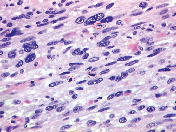
Figure 8:
Leiomyosarcoma comprised of pleomorphic spindle cells with mitotic activity
(encircled) [12].
Case 2: A
38-year-old man had manifested with multiple lesions over his penis of 8 months
duration. His clinical examination showed a fungating mass over his glans penis
that had measured 3 cm x 4 cm and which had involved his external urethral
meatus and which was associated with three separate mobile nodules over the
proximal shaft of his penis (see Figure
9). There was no evidence of inguinal lymph node enlargement. He had punch
biopsy of the penile lesion and pathology examination of the specimen had shown
high-grade leiomyosarcoma of penis. Immunohistochemistry staining studies of
the tumor did show that the tumor cells had exhibited positive staining for SMA
as well as desmin. The tumor cells did exhibit negative staining for S-100,
CD34 and Myogenin. He did undergo total penectomy with perineal urethrostomy.
In view of the fact that there was no evidence of inguinal lymph node
enlargement, lymph node dissection was not undertaken.
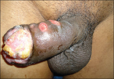
Figure 9:
Clinical photograph [14].
He was well at his 9 months follow-up with no
evidence of local recurrence or distant metastasis. There is no consensus
opinion regarding the best treatment options for primary leiomyosarcoma of the
penis. Generally many clinicians would be happy with radical excision of tumor
by total penectomy and perineal urethrostomy ensuring complete excision of the
tumor alone to be followed by careful follow-up of patients at regular
intervals with clinical and radiology imaging studies. Others would recommend
adjuvant therapy (chemotherapy alone, radiotherapy alone or radiotherapy plus
chemotherapy). Other clinicians would recommend entering such patients into an immunotherapy trial.
Considering the fact the case was reported with 9
months follow-up one cannot predict what the long term outcome of the patient
would be. Dabernig, et al. reported a case of leiomyosarcoma of the penis in
which the surgical treatment had involved sub-cutaneous penectomy with the
preservation of a sensate skin envelope, bilateral groin dissection, and
perineal urethrostomy [15]. Reconstruction of the
urethra and soft tissue was undertaken with utilization of a free radial
forearm flap. Dabernig, et al. have the opinion that subcutaneous penectomy
should be considered as a treatment option in selected cases of penile tumor in
view of the fact that the procedure does facilitate urethral reconstruction [15].
Conclusions
· Primary
leiomyosarcomas of the penis are rare malignancies that mimic other lesions of
the penis therefore careful histopathology and immunohistochemistry examination
is required to confirm its diagnosis.
· Most
cases of superficial primary leiomyosarcomas of the penis (SLPOPs) would tend
to have good outcome following complete surgical excision of the lesions but
very large superficial tumors with high mitotic activity could subsequently
recur and they should be followed up carefully.
· Deep
leiomyosarcomas of the penis do have a tendency to recur and some of these tumors
have been treated by means of radical excision alone, radical excision plus
radiotherapy, radical excision plus both chemotherapy and radiotherapy but
controversies exist regarding the usefulness of adjuvant
chemotherapy/radiotherapy in the treatment of deep-seated primary
leiomyosarcomas of the penis that are not associated with lymph node
enlargement or metastasis (localized deep seated primary leiomyosarcomas of the
penis).
· There
is need for the establishment of a global multi-center trial of the utilization
of immunotherapy, chemotherapy and radiotherapy in the treatment of deep
leiomyosarcomas of the penis in order to streamline treatment guidelines for
the disease.
Acknowledgement
Indian Journal of Cancer and Wolters Kluwer Medknow publications
for granting permission for reproduction of contents and figures from their journal
under copyright.
This article is available under the terms of the
Creative Commons Attribution-Non Commercial-Share Alike License (CC BY-NC-SA),
which permits non-commercial use, distribution and reproduction in any medium
provided the original work is properly cited. Urology Case Reports for granting
copyright permission for reproducing contents and figures from there Journal
under copyright © 2015 The Authors this is an open access article under the CC
BY-NC-ND license.
(http://creativecommons.org/licenses/by-nc-nd/4.0/).
Sarcoma and Hindawi Publishing Corporation for
granting permission for reproduction of contents and figures from their journal
article under copyright © 2002 Hindawi Publishing Corporation. This is an open
access article distributed under the Creative Commons Attribution License,
which permits unrestricted use, distribution, and reproduction in any medium,
provided the original work is properly cited.
Indian Journal of Pathology and Microbiology and
Wolters Kluwer Medknow Publications for granting permission for reproduction of
contents and figures from their Journal article under copyright © 2009, Wolters
Kluwer Medknow Publications. This article is available under the terms of the
Creative Commons Attribution-Non Commercial-Share Alike License (CC BY-NC-SA),
which permits non-commercial use, distribution, and reproduction in any medium,
provided the original work is properly cited.
References
1. Fetsch JF, Davis CJ, Miettinen M and Sesterhenn
IA. Leiomyosarcoma of the penis: a clinicopathologic study of 14 cases with
review of the literature and discussion of the differential diagnosis (2004) Am
J Surg Pathol 28: 115-125. https://doi.org/10.1097/00000478-200401000-00014
2. Sundersingh S, Majhi U, Narayanaswamy K and
Balasubromanian S. Primary leiomyosarcoma of the penis (2009) Indian J Pathol
Microbiol 52: 447-448. https://doi.org/10.4103/0377-4929.55028
3. Chaux
A and Cubilla AL. Penis and Scrotum other malignancies Leiomyosarcoma (2010) PathologyOutlines.com,
Inc., USA.
4. Pow-Sang MR and Orihuela E.
Leiomyosarcoma of the penis (1994) J Urol 151: 1643-1645. https://doi.org/10.1016/s0022-5347(17)35328-4
5. Nanri M, Kondo T, Okuda H, Tanabe K and Toma
H. A case of leiomyosarcoma of the penis (2006) Int J Uro 13: 655-658. https://doi.org/10.1111/j.1442-2042.2006.01376.x
6. Kathuria S, Jablokow VR and Molnar Z.
Leiomyosarcoma of the penile prepuce with ultrastructural study (1986) Urol 27:
556-557. https://doi.org/10.1016/0090-4295(86)90345-6
7. Dominici A, Delle Rose A, Stomaci N,
Pugliese L, Posti A, et al. A rare case of leiomyosarcoma of the penis with a
reappraisal of the literature (2004) Int J Urol 11: 440-444. https://doi.org/10.1111/j.1442-2042.2004.00806.x
8. Katsikas VS, Kalyvas KD, Ioannidis SS,
Papathanasiou MV, Panagiotopoulou KP, et al. Leiomyosarcoma of the penis (2002)
Sarcoma 6: 75-77. https://doi.org/10.1080/1357714021000022177
9. Pratt RM and Ross RT. Leiomyosarcoma of
the penis. A report of a case (1969) Br. J Surg 56: 870-872. https://doi.org/10.1002/bjs.1800561122
10. Greenwood
N, Fox H and Edwards EC. Leiomyosarcoma of the penis (1972) Cancer 29: 481-483.
Primary, Leiomyosarcoma, Penis, Superficial,
Deep, Wide excision, Partial penectomy, Total penectomy, Radiotherapy, Chemotherapy,
Desmin, Smooth muscle actin, Spindle cells, Mitoses, Atypia ">
Primary, Leiomyosarcoma, Penis, Superficial,
Deep, Wide excision, Partial penectomy, Total penectomy, Radiotherapy, Chemotherapy,
Desmin, Smooth muscle actin, Spindle cells, Mitoses, Atypia