Background/Purpose: Agenesis is an anomaly where the tooth germ
fails to differentiate completely into dental tissues resulting in congenitally
missing teeth. This is one of the commonest dental anomalies. The aim of this
study was to determine the prevalence of bilateral agenesis of maxillary
lateral incisors. This study also reflects upon the treatment options available
when there is agenesis of maxillary lateral incisors. Materials and Methods: Bilateral agenesis was considered and
included in the study as agenesis and unilateral agenesis was excluded from the
study. Orthopantamograms (OPGs) of 945 dental patients aged 6-30 years were
examined for the agenesis of teeth. Frequencies were calculated using chi
square test and the level of significance was considered if p value was
<0.05. Results: The prevalence of
bilateral agenesis or congenitally missing maxillary lateral incisors was at
8.2%. Conclusions: The prevalence rate
of bilateral agenesis of maxillary lateral incisors is more common in females
than males. An evidence based decision should be taken for the clinical
management of this kind of agenesis.
Introduction
Agenesis
or congenitally missing tooth occurs when the tooth germs fail to differentiate
appropriately into dental tissues [1,2]. Agenesis has shown a high prevalence
amongst the population at around 25% [3,4]. Agenesis when seen in less than six
teeth (excluding third molars) is defined as Hypodontia
[4]. Anodontia refers to condition when there is complete agenesis of teeth and
Oligodontia is defined as the condition when there is agenesis of six or more
teeth and the term [5,6].
Agenesis
is commonly seen in third molars and after the third molars agenesis is seen
more commonly with mandibular second premolars and then the maxillary lateral
incisors [7,8]. There can be arch length discrepancies, malocclusion
and unaesthetic appearance as a result of agenesis [9,10]. Several etiological
factors have been suggested for the development failure of the permanent tooth
germ, thus leading to its absence, such as: physical obstruction, dental lamina
rupture, limitation of space or functional anomalies [11]. In spite of recent
progress, the etiopathogenesis of hypodontia remains largely unknown. There is
evidence that congenital
tooth absence can be the result of environmental or
hereditary causes, or even of their interaction.
Factors
like genetics and dietary factors have been suggested as responsible for the
etiology of agenesis of teeth [11,12]. When the primary and permanent
dentitions have been compared it is seen that the permanent dentition has
increased prevalence of agenesis when compared with primary dentition [13]. Orthodontic
treatments can be affected when there is agenesis
of maxillary
lateral incisors.
When
the agenesis or congenitally missing teeth is in the functional, esthetic or
more anterior region it can have an imminent psychological and functional ill
effect on the patient [12]. It has been emphasized that early diagnosis of
hypodontia can result in minimal functional, psychological and esthetic
complications which may have to be dealt with later in life of the patient
[12,14]. Orthodontic space re-distribution, fixed
partial denture and implants are considered as standard
treatment options for these patients which can help the patient lead a normal
functional life [14-16].
The
current study was designed to understand the prevalence of bilateral agenesis
of maxillary lateral incisors. The authors also have tried to suggest possible
clinical management options of congenitally missing maxillary lateral incisors.
Materials and Methodology
This
was a retrospective, observational study conducted after approval from the
Research and Ethics Committee at RAK
College of Dental Sciences (RAKCODS), RAK
Medical and Health Sciences University (RAKMHSU),
RAK, UAE. The objective of this study was to understand the prevalence of
bilateral agenesis of maxillary lateral incisors. The study also intended to
evaluate the gender and arch predilection for the bilateral agenesis of
maxillary lateral incisors. The age group of the patients of whom the OPGs were
selected was between 6 years to 30 years of age. OPGs which showed bilateral
agenesis of maxillary lateral incisors were included. Since a clinical examination
of these patients was not possible only those OPGs which showed bilateral
absence were considered to be true agenesis and were included in the present
study.
945
Orthopantamograms (OPGs) were first included out of the total 18500 OPGs
available. The electronic health files of patients were evaluated to exclude
patients with syndromes. Within these 945 OPGs bilateral agenesis was sought
for. The sampling followed in this study was convenience sampling as the team
selected the OPGs which fit into the selection criteria.
Statistical Analysis
Data
observed in this study was described using descriptive statistical analysis. To
evaluate the frequency of agenesis between the sexes (males/females),
chi-square statistical test was applied, the level of significance was set at
P<0.05.
Results
Bilateral
agenesis was found in 85 OPGs out of the 945 OPGs. 8.2% showed bilateral
agenesis or congenitally missing maxillary lateral incisors (The remaining of
these 85 OPGs showed bilateral agenesis of mandibular
second premolars and third molars) (Figure 1).
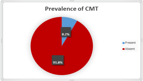
Figure 1: Prevalence
percentage of bilateral agenesis of maxillary lateral incisors.
The
prevalence of bilateral agenesis or congenitally missing teeth was seen more in
females (57.14%) than in males (42.85%) (Table
1), the results were however not statistically significant (X2=0.98,
P=0.26).
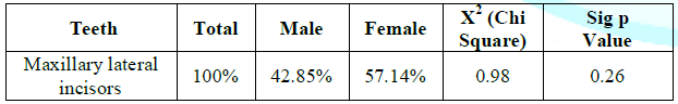
Table 1: Prevalence of
bilateral agenesis of maxillary lateral incisors between males and females.
Discussion
Studies
have suggested agenesis to have a prevalence rate of 25% amongst the general
population making it one of the commonest dental
anomalies in humans [9]. This anomaly is
associated with other conditions like crowding and delayed eruption [10]. The permanent
dentition is more affected with agenesis when
compared to the primary dentition [11]. In the present study the prevalence
rate of bilateral agenesis of maxillary lateral incisors has been evaluated.
In
this retrospective study a total of 945 OPGs were initially included of which
85 OPGs showed evidence of bilateral agenesis or congenitally absent teeth
including third molars. Out of these 85 OPGs 8.2% reflected agenesis of
maxillary lateral incisors.
Gender predilection
The
prevalence for bilateral agenesis of maxillary lateral incisors was seen more
in females (57.14%) than males (42.85%) (Table 1). There are studies which have
shown results similar to the present study where there is an increased rate of
prevalence of agenesis in females when compared to males [13,17]. It has been
shown in studies that the prevalence of hypodontia is usually higher in females
[10]. Other studies have showed higher incidence rates in males when compared
to females [12,18,19]. However when the literature is explored there is no much
evidence or reasoning as to why the prevalence is higher or lower in either
gender though genetics and hereditary factors have been attributed as a strong
reasoning factor [20].
Clinical Management of Agenesis Related to Maxillary
Lateral Incisors
The
management of bilateral agenesis or congenitally missing lateral incisors can
be divided into the following scenarios
·
When there is space between the
maxillary central incisors and canines
·
When there is no space between
the maxillary central incisors and canines
When there is space between the maxillary central
incisors and canines
Whenever
the case presents with adequate space (Appropriate mesiodistal width) between
the maxillary central and canine depicting the actual space available for a
lateral incisor, then the best treatment will be a single tooth
implant restoration. However factors like
orthodontic redistribution of space may need to be considered dependent upon
the case. Earlier on options like resin bonded bridge or a fixed partial
denture was also used successfully. However these treatment options had their
own drawbacks like the possibility of endodontic
treatment needed for the abutment teeth later on
or the possibility of pulpal injury if no intentional endodontic treatment was
done before the bridge placement. However now single tooth implant restoration
looks to be the choice of treatment [20,21].
When there is no space between the maxillary central
incisors and canines
Whenever
the case presents with no adequate space between the maxillary central incisor
and canine, the canine is more or less in the space of the lateral incisor. In
these scenarios the main concern is regarding the appearance of the canines.
The options available include reshaping the canines to the shape of laterals.
If the reshaping is not esthetic enough then the option of laminates or veneers
can be explored upon. However the thickness of canines reduction need to be
considered for a replacement with veneers or laminates as the tooth will
require more reduction considering the bulk of the canine. In these cases the
possibility of intentional endodontic treatment of the canines is a definite
possibility to look into before the placement of laminates or veneers [20,21].
The
authors of the present study believe that there should be increased number of
samples included to give formidable results with regards to the prevalence of
bilateral agenesis of maxillary lateral incisors and also to understand the
gender predilection clearly regarding the prevalence rates. Studies should also
be done on a larger scale to understand the genetics behind agenesis. However
the management options of agenesis of maxillary lateral incisors can get
challenging at times and any treatment planned should be well thought as the
patient will have to live with it for a lifetime. It has to be understood that
there are no straightforward methods in the management of agenesis of the
maxillary lateral incisors but the dentist has to be more flexible,
accommodative and actually innovative in understanding the situation comprehensively
and deal accordingly.
Conclusions
In
the present study we found that
· The
prevalence rate of bilateral agenesis or congenitally missing maxillary lateral
incisors is at 8.2%.
· Clinical
management options of congenitally missing maxillary lateral incisors need to
be based on evidence based dental practice.
References
- Moyers RE and Riolo ML.
Handbook of orthodontics, Moyers RE (Ed) (1988) Year Book Medical Publishers,
USA, pg-348‑53.
- Silva MR. Radiographic
assessment of congenitally missing teeth in orthodontic patients (2003) Int J
Paediatr Dent 13: 112‑116.
https://doi.org/10.1046/j.1365-263x.2003.00436.x
- Rakhshan V. Meta-analysis
and systemic review of factors biasing the observed prevalence of congenitally
missing teeth in permanent dentition excluding third molars (2013) Prog
Orthod14: 33.
- https://doi.org/10.1186/2196-1042-14-33
- Bural C, Oztas E, Ozturk
S and Bayraktar G. Multidisciplinary treatment of nonsyndromic oligodontia
(2012) Eur J Dent 6: 218-216. https://doi.org/10.1055/s-0039-1698954
- Closs LQ, Weissbluth MF,
Nakamura E and Hermann FP. Esthetic and functional rehabilitation for
oligodontia in the mixed dentition: case report (2012) J Dent Child 79: 193-196.
- Parkin N, Elcock C, Smith
RN, Griffin RC and Brook AH. The aetiology of hypodontia: the prevalence,
severity and location of hypodontia within families (2009) Arch Oral Biol 54:
52-56.
https://doi.org/10.1016/j.archoralbio.2008.11.002
- Lo Muzio L, Mignogna MD,
Bucci P and Sorrentino F. Statistical survey on the incidence of agenesis in a
sample of 1529 subjects (1989) Minerva Stomatol 28: 1045-1051.
- Laganà G, Venza N,
Borzabadi-Farahani A, Fabi F, Danesi C, et al. Dental anomalies: prevalence and
associations between them in a large sample of non-orthodontic subjects, a
cross-sectional study (2017) BMC Oral Health 17: 62.
https://doi.org/10.1186/s12903-017-0352-y
- Kokich VO Jr and Kinzer
GA. Managing congenitally missing lateral incisors Part II: Tooth-supported
restorations (2005) J Esthet Restor Dent 17: 76-84.
https://doi.org/10.1111/j.1708-8240.2005.tb00089.x
- Abu-Hussein M, Watted N,
Azzaldeen A, Yehia M and Awadi O. Prevalence of Missing Lateral Incisor
Agenesis in an Orthodontic Arabs Population in Israel (Arab48) (2015) Int J Pub
Heal Res 3: 101-107.
- Larmour C, Mossey PA,
Thind BS, Forgie AH and Stirrups DR. Hypodontia-A retrospective review of
prevalence and etiology. Part I (2005) Quintessence Int 36: 263-270.
- Vahid-Dastjerdi E,
Borzabadi-Farahani A, Mahdian M and Amini N. Non-syndromic hypodontia in an
Iranian orthodontic population (2010) J of Oral Sci 52: 455-461.
https://doi.org/10.2334/josnusd.52.455
- Polder BJ, Van’t Hof MA,
Van der Linden FP and Kuijpers-Jagtman AM. A meta-analysis of the prevalence of
dental agenesis of permanent teeth (2004) Comm Dent Oral Epidemiol 32: 217-226. https://doi.org/10.1111/j.1600-0528.2004.00158.x
- Kokich VG and Kokich VO.
Congenitally missing mandibular second premolars: Clinical options (2006) Am J
Orthod Dentofacial Orthop 130: 437-444.
https://doi.org/10.1016/j.ajodo.2006.05.025
- Kokich VO Jr and Kinzer
GA. Managing congenitally missing lateral incisors Part II: Tooth-supported
restorations (2005) J Esthet Restor Dent 17: 76-84.
https://doi.org/10.1111/j.1708-8240.2005.tb00089.x
- Kennedy D. Missing second
premolars: can early treatment make a difference? (2010) Spring Vancouver
Canada: PCSO Bulletin pg 29-33.
- Spear F, Mathews D and
Kokich V. Interdisciplinary management of single-tooth implants (1997) Semin
Orthod 3: 45-72.
- Gracco ALT, Zanatta S,
Valvecchi FF, Bignotti D, Perri A, et al. Prevalence of dental agenesis in a
sample of Italian orthodontic patients: an epidemiological study progress in
Orthodontics (2017) Prog Orthod 18: 33. https://dx.doi.org/10.1186%2Fs40510-017-0186-9
- Ali S, Al Hur AA,
Alyessary AA and Ali G. Prevalence of congenital missing permanent teeth in a
sample of Iraqi patients attending dental clinics of Kerbala University: A
Retrospective Study (2019) Biochem Cell Arch 19: 3265-3272.
- Millar BJ and Taylor NG.
Lateral thinking: the management of missing upper lateral incisors (1995) Brit
Dent J 179: 99-106.
- Miller TE. Implications
of congenitally missing teeth: orthodontic and restorative procedures in the
adult patient (1995) J Pros Denti 73: 115-122. https://doi.org/10.1016/s0022-3913(05)80148-9
*Corresponding author
Vivek Padmanabhan, Assistant
Professor, Pediatric and Preventive Dentistry Department, RAK College of Dental
Sciences, RAK Medical and Health Sciences University, United Arab Emirates, E-mail:
vivek.padmanabhan@rakmhsu.ac.ae
Citation
Padmanabhan V, Omar Khaled AM and Rahhal LMK. Prevalence
of bilateral agenesis of maxillary lateral incisors and clinical management
options (2020) Dental Res Manag 4: 31-33.
Maxillary lateral incisors, Orthopantamograms,
Bilateral agenesis.


