

Case Report :
Yuan-Chieh Chang,
Ming-Ke Tsai and Jian-Hong Yu A
20-year-old man presented at our clinic with a primary complaint of irregular
dentition and reverse bite. Clinical examination revealed skeletal Class III
malocclusion with an anterior crossbite and a completely blocked #25 in the
maxilla. Because he refused to receive orthognathic surgery and experienced
only mild functional interference, a nonextraction treatment method was
adopted. Space creation on the left upper arch was successfully performed using
an open coil spring for molar distalization and the improved super-elastic
Ti–Ni alloy wire (ISW) Multi-bends Edgewise Arch Wire (MEAW) technique.
Furthermore, #25 was aligned completely. The Class III malocclusion was
corrected using the ISW MEAW technique combined with Class III elastics on the
lower arch. Treatment was completed in approximately 16 months and a
satisfactory occlusion was achieved after active treatment. An improved super-elastic Ti–Ni alloy
wire (ISW) was developed by Tokyo Medical and Dental University and marketed
under the brand name L&H® Titan wire. Low-hysteresis wires deliver
relatively stable orthodontic force in oral cavities [1]. In
contrast to the hardness and stiffness of stainless steel wires, ISW offers
three main advantages, namely shape memory, super elasticity, and absorption of
shock and vibration [2]. Thus, individual occlusion through orthodontic treatment
can be easily achieved using the ISW technique. Figure
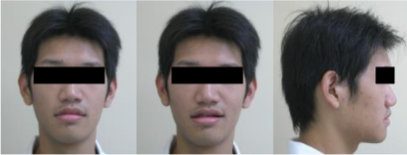
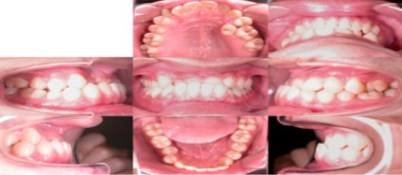
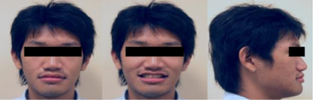
1: Facial photographs before treatment. Figure
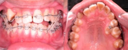
2: Intraoral photographs before treatment. A 20-year-old man arrived at the
dental clinic presenting with occlusion of his anterior teeth (Figure 1 and Figure 2). The Angle
classification was Class III malocclusion. Furthermore, tooth #25 was blocked
palatally in the upper left dental arch. At the anterior occlusion, the patients
upper and lower incisors exhibited edge-to-edge contact. This case report
provides details of ISW application for the treatment of the Class III
malocclusion with anterior crossbite dentition. The patient had no history of systemic diseases. He
had two impacted third
molars, specifically in teeth #38 and #48 (Figure 3). Figure
3: Panoramic film before treatment. The following treatment plans
were established: (1) Extraction of teeth #38 and #48; (2) Full-mouth direct
bonding through bracket and ISW application; (3) Placing the crossbite arch between
#13 and #23; and (4) Bringing #25 into alignment. We initially extracted teeth #38
and #48. Next, we applied ISW with a crossbite arch between #13 and #23, thus
forming a loop between #11 and #21 for anterior crossbite correction. A 100-gf
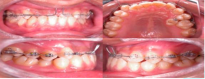
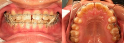
open coil spring was placed between #24 and #26 to create space for #25 (Figure 4). Figure
4: 0 weeks of active treatment. After 2 weeks of active
treatment, the anterior teeth formed edge-to edge contact. The ISW Multi-bends
Edgewise Arch Wire (MEAW) [3] was applied on the upper right dentition for
additional space creation (Figure 5).
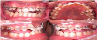
Anterior crossbite was corrected using the ISW crossbite arch technique.
Meanwhile, the distalization of #27 was performed and Class III intermaxillary
elastics were used to prevent the lower anterior teeth
from flaring out (Figure 6). Figure 5: 2 weeks of active treatment. Figure
6: 3.5 Months of active treatment. After 9.5 months, #26 moved
distally because of the use of the stopper and 150-gf open coil spring. Simultaneously,
#25 was tied with the ligature wire to lightly guide its movement to the buccal
side (Figure 7). Figure
7: 9.5 Months of active treatment. After 16 months of active
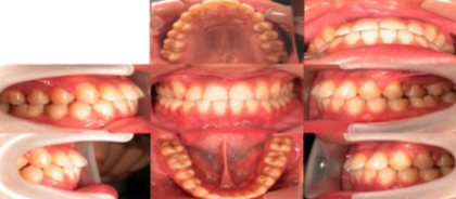
treatment, satisfactory occlusion was achieved (Figure 8 and Figure 9). Figure
8: Facial photographs after treatment. Figure
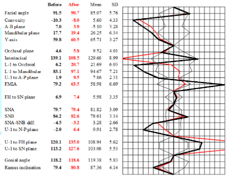
9: Intraoral photographs after treatment. Cephalometric analysis
results and superimposition revealed that the upper incisors were
tipping outside between 120.1° and 135.0° on the
Frankfurt horizontal plane (Figure 10
and Figure 11). The mandibular plane angle increased from 17.7°
to 19.4°. Figure
10: Cephalometric analysis. Black and red
lines indicate positions before and after treatment, respectively In 1988, Graber defined crossbite
dentition as a malocclusion in which one or more teeth might be abnormally
positioned in reference to the opposing teeth. The reported incidence of
anterior dental crossbite is 4%-5% [4]. The causes of reduced anterior crossbite
dentition are inadequate arch length, trauma, a repaired cleft lip, an
over-retained deciduous tooth, or length discrepancy between the maxilla and
mandible [5]. In 1981, Rakosi and Schilli also described some of the effects of
environmental factors, such as mouth-breathing and oral habits, on the etiology
of Class III malocclusion. A removable acrylic plane can be used during orthodontic treatment
of anterior crossbite cases of early mixed dentition [6]. Furthermore, a palatal plate with
anterior springs or a quad-helix appliance to which springs can be soldered can
be considered if patients are willing. Deciding the treatment for Class
III cases is always challenging in orthodontics because
it is difficult and time consuming [7]. The key to achieving successful
treatment outcomes lies in accurate differential diagnosis of a Class III case
as a dental functional or skeletal problem. At the beginning of active
treatment, we discovered that our patient exhibited a true skeletal Class III
problem according to the results of a cephalometric analysis;
however, clinical examination of the edge-to-edge contact of the anterior teeth
revealed mild functional interference around the anterior teeth. Therefore, the
crossbite dentition could be corrected without surgery. After 16 months of
active orthodontic
treatment, crowding was relieved and an adequate overbite and over jet were
attained. Previously, the crossbite arch dentition
could not be easily corrected because treatment of anterior crossbite usually
required a lingual arch
with finger-like springs and a bite plane [8]. Moreover, available treatments
that required a lingual arch with springs were associated with certain
disadvantages. First, torque or space control was limited, and designing
orthodontic appliances based on the force of the mechanism was difficult.
Second, the appliances could injure the temporomandibular joint
and induce some problems while using the bite plane [5]. Third, additional
effort and time was sometimes required to prepare an acrylic resin or block for
the orthodontists to apply on patients. In summary, an efficient and simple
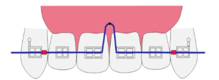
design for treating patients with crossbite arches is necessary. ISW crossbite
arches are easily bent and do not hurt the joints because they do not increase
the occlusal height (Figure 12). Most importantly, the degree of
correction can be estimated in advance by altering the position of stoppers;
furthermore, correcting crossbite requires less time and can be performed
immediately at dental clinics. 2.
Garrec P, Tavernier B and Jordan
L. Evolution of flexural rigidity according to the cross-sectional dimension of
a superelastic nickel titanium orthodontic wire (2005) Eur J Orthod 27: 402-407. https://doi.org/10.1093/ejo/cji014 3.
Beane RA Jr. Nonsurgical
management of the anterior open bite: a review of the options (1999) Semin
Orthod 5: 275-283. https://doi.org/10.1016/S1073-8746(99)80021-8
4.
Major PW and Glover K. Treatment
of anterior cross-bites in the early mixed dentition (1992) J Can Dent Assoc 58:
574-575, 578-579. 5.
Prakash P and Durgesh BH.
Anterior Crossbite Correction in Early Mixed Dentition Period Using Catlans
Appliance: A Case Report (2011) ISRN Dent 298931. https://dx.doi.org/10.5402%2F2011%2F298931
6.
Jirgensone I, Liepa A and
Abeltins A. Anterior crossbite correction in primary and mixed dentition with
removable inclined plane (Bruckl appliance) (2008) Stomatologija 10: 140-144. 7.
Thalanki LP. Nonsurgical treatment
of Class III malocclusion (2007) J Mass Dent Soc 56: 40. Jian-Hong Yu, Professor,
School of Dentistry, College of Dentistry, and Dean, Department of Orthodontics,
China Medical University Hospital, Taichung-40402, Taiwan, R.O.C. E-mail: kenkoyu@mail.cmu.edu.tw Chang YC, Tsai MK and Yu JH. Improved
super-elastic Ti–Ni alloy wire treatment for skeletal class III malocclusion with
anterior crossbite dentition (2019) Dental Res Manag 3: 6-8. Super-elastic
Ti–Ni alloy, Orthodontic treatment, Crossbite dentition and Cephalometric
analysisImproved Super-elastic Ti–Ni Alloy Wire Treatment for Skeletal Class III Malocclusion with Anterior Crossbite Dentition
Abstract
Full-Text
Introduction
History
Treatment Plan
and Progress
Discussion
References
1.
Liaw YC, Su YY, Lai YL and Lee
SY. Stiffness and frictional resistance of a superelastic nickel-titanium
orthodontic wire with low-stress hysteresis (2007) Am J Orthod Dentofacial
Orthop 13: 512-578. https://doi.org/10.1016/j.ajodo.2006.08.015
*Corresponding author
Citation
Keywords

