

Introduction
Everyone agrees that a good understanding of occlusion is essential to ensure optimum dental health. Unfortunately, that seems to be the only point of consensus for this important, yet controversial subject. Occlusion is often described as troublesome, complex, and incoherent, but confusing seems to describe it best.
Christensen comments: Unfortunately, occlusion in its broad definitions is not a popular subject in CE or in actual practice. Although occlusion principles permeate almost all of dentistry, the area is confounded by confusing theories, non-practical techniques, contradictory beliefs, and practitioners unaware of the basic concepts of occlusion. As a result, most dental patients go without the benefits of dental therapy based on several occlusal principles [1].
The confusion has left us with many unanswered questions, the most important being, the precise role that occlusion plays in the etiology of Temporo Mandibular Joint Disorders (TMJD). Since it is unknown, we have to consider the possibility that the way we are doing our work, may be a contributing factor. The agenda for this commentary is in three parts. The first part will thoroughly discuss the three factors that have largely contributed to this abstruseness. The second will be dedicated to Para function, and the third will review how dental work is being performed today, and will conclude with guidelines for the restorative dentist that will minimize and prevent problems with the Temporo Mandibular Joint (TMJ).
Occlusion Confusion
Three
Sources of Confusion
Definition
How many times have you heard of
a restorative failure being blamed on the occlusion? What exactly does that
mean? The question is ambiguous because the word occlusion has three different
interpretations. The original definition was the act or process of occluding
from the Latin, occludere, which means to shut up or close up [2]. Dental
occlusion therefore would be defined as the
relation of the teeth when the jaws are closed nothing more. This was 50 years
ago.
But today, there are two additional meanings. Dorlands Pocket Medical Dictionary first defined dental occlusion correctly as‚ the closure of teeth, but later expanded the definition to the contact of the teeth of both jaws during those excursive movements of the mandible essential to the function of mastication. [3] Why did they do this when early studies suggested that; 1. Teeth rarely and lightly touch during mastication [4] and 2. Excursive movements of the mandible are not essential to the function of mastication? [5]
The dental profession had witnessed patients grinding their teeth in lateral excursions, saw that it was doing damage, and sought to find the most comfortable pathway, i.e., the horizontal vector with the least resistance or interference for them to do so. Many assumed that these horizontal excursions were an integral part of normal function, when in reality it is parafunction. People do not normally eat in horizontal excursions unless forced to do so by a flattened dentition.
Still another description over the years, the word occlusion morphed into a homonym for the stomatognathic system. Jablonskis definition of occlusion is a sixty word description of all the components of the stomatognathic system. [6]. It is not a valid definition of occlusion. The different interpretations are a major distraction. It clouds the issues and makes questions such as, what has occlusion got to do with TMD? impossible to answer. Are we asking if tooth contact during closure is causing a problem with the TMJ, or do we want to know if the stomatognathic system is not functioning properly?
Modern day texts on occlusion do not just describe the simple touching of upper and lower teeth, but rather present a detailed analysis of the whole stomatognathic system. Occlusion (the way teeth touch each other during closure) and the stomatognathic system are two distinct entities and should be described separately to remove the present ambiguity.
Function
of the Stomatognathic System: Two Different Views
Without a doubt, the biggest
source of confusion comes from the fact that there are two entirely different
viewpoints as to how the stomatognathic system should function.
The Vertical Function Paradigm: In this model, the mandible functions vertically. We talk vertically, we swallow vertically, and we eat vertically. The vector of mastication is a vertical teardrop with a lateral movement of 5mm to 6 mm during the first phase of chewing, and as the teeth approach each other, the lateral displacement lessens to 3mm to 4mm from the starting position. The mandible is guided into position during closure by the occlusal incline planes of the teeth. It is not a consistent and reproducible movement, but is a function of head position. As the head tilts forward, the mandible goes forward. After each vertical function, the mandible returns to a state of physiological rest [7].
During opening and closing, there is condylar centricity where the axis is maintained, and upon complete closure, the condyles are seated in the anterior-superior portion of the glenoid fossae. Border movements are seldom used and most mandibular movements take place within a reasonably limited three dimensional space. The model in this paradigm is free from parafunction.
The Horizontal Function Paradigm: While there is no disagreement that we talk and swallow vertically, the horizontal paradigm is focused on the premise that the mandible functions laterally, rather than vertically. How did this come about?
In the early 20th century there were a number of dentists who were preoccupied with the mandibles ability to rotate around axes in three planes. The study of these jaw movements became known as gnathology, and its followers gnathologists. It was the objective of the gnathologists to produce a proper occlusal form which was dictated by the determinants of mandibular movements, i.e., to create an occlusal surface that would accommodate free passage for the opposing dentition.
Their goal was to eliminate interferences in laterotrusive movements from centric. It was called optimal functional occlusion [8]. But theres a conflict. Functional occlusion is defined as the touching of upper and lower teeth during mastication and deglutition-both are vertical not horizontal. So the gnathologists had the perception that it is normal to function in lateral excursions, but the reality is that it is parafunction.
The gnathologists had another conflict: the anterior teeth which are a major interference to anterior excursions. If they were to eliminate that obstacle, they would have to shorten the incisors considerably. Now what? Their solution was to make lemonade. They declared that that purpose of the anterior teeth being longer was to disengage the posterior teeth during these parafunctional excursions for their protection. It was called anterior guidance. Nothing was said about the trauma that the anterior teeth would receive during this exercise.
Discussion
So on the one hand, we have a paradigm that describes a system that functions vertically, pays little attention to border movements, and is free from parafunction. On the other, we have a paradigm whose goal is to eliminate interferences in laterotrusive movements. Which is best? The vertical paradigm is the model we want to emulate as it is free from parafunction. If parafunction does occur, treatment should focus on management and prevention, not on accommodation.
The Morphology of Teeth
There are three key benchmarks that are used when evaluating the overall status of the stomatognathic system: the condyle, the muscles of mastication, and the dentition. There is no disagreement that when the system is healthy and functioning efficiently, the muscles of mastication are relaxed and the condyles are seated properly in the anterior superior part of the fossae. There are however, major questions regarding the dentition. There is no consensus regarding their morphology, how exactly they should touch each other in closure, and more importantly, their involvement in TMJ disorders.
From an engineering point of view, what is the best design for teeth that allows the system to function efficiently?
There are design principles that appear to govern the structure-function relationship in organisms, i.e., there is an interface between mechanical engineering and biology. The idea being that biological materials and structures are designed for specific functions [9]. In regard to teeth, the original architect gave us a perfect example. The purpose of the fossae is to hold food for cutting, while the sharp cusps have two purposes: to cut the food and to direct mastication forces vertically down the long axis upon closure.
The space between the incline planes allows resistance-free repositioning of the mandible/condyle not only during swallowing, but anterior posterior postural changes as well. So it would appear that the original design or morphology of our teeth is best suited to serve our stomatognathic system. But unfortunately, this is not the standard that everyone emulates.
For instance, when constructing removable dentures, we have a choice of seven different designs for the posterior teeth-from 33 degrees to flat plane occlusion. Now, why would we want to provide flat plane occlusion for a 90 year old denture patient with no alveolar ridge, who has a diminished mastication force of 75% forcing that patient to mash his/her food laterally, distracting the condyles, and dislodging the dentures?
What were the circumstances that programmed us for the concept of flat plane occlusion? There were three: one was the influence of the horizontal function paradigm, but the strongest influence was some research that was done by Kydd, Regli, Swoope, and Ortman in the 1950s and 60s. [10-13]. These investigators placed strain gauges in dentures and had patients eat various types of foods with different morphologies of posterior teeth from 33 degrees to zero degree. The results: the strain gauges measured highest with 33 degree teeth and lowest with the zero degree. Their conclusions were that the increased strain in the denture with 33 degree teeth would be harmful to the alveolar bone. Unfortunately, their conclusions lacked understanding. The fact that the strain gauges registered high with the 33 degree teeth meant that the denture was working efficiently, directing valuable vertical stimulation to the alveolar ridge.
The zero degree teeth diminished the alveolar stimulation, dislodging the dentures laterally as the flattened teeth forced the patient to eat laterally. And finally there is the perception that our evolutionary blueprint has programmed us for zero degree occlusion and that all living humans were designed to eat with a flattened dentition. Neilburger (dentist and anthropologist) warns that deviation from this model may cause serious problems for patients and he encourages dentists to aid in this attrition [14]. He labels this process as normal. It is normal because it is common, but common isnt necessarily good. It is poor speculation to declare that we are predestined to have a flattened dentition as evidenced by the many seniors who maintain naturally sharp teeth.
Dental Compression Syndrome
Disordered
or Perverted Function
The
Common Denominator for the Sources of Confusion
McCollum and Stuart once talked
about a subtle pathology of function between the opposing teeth and movements
of the mandible. They declared that the lack of understanding regarding this
pathology has kept dentistry from the opportunity to render substantial health
services to our patients. That subtle pathology is Dental
Compression Syndrome (DCS), aka Parafunction, aka
Bruxism. DCS is defined as a total parafunctional daily or nightly activity
that includes grinding, gnashing, or clenching of ones teeth. Capable of forces
in excess of 500 pounds per square inch, DCS can inflict compressive, tensil,
shearing, and flexural forces on the dentition while simultaneously imposing
unwanted force to the alveolar
bone and the TMJ. Our goals in restorative
dentistry are twofold; besides successfully completing the current project,
whether it is a crown, alloy restoration, or denture, it is also our obligation
to keep the patients stomatognathic system healthy and comfortable until the
next restorative project - the biggest threat to that goal is DCS.
Signs of Dysfunction
One reason DCS has been so successful over the centuries is that it works well within ones subconscious. Since many patients affected are unaware, the General Practitioner GP must recognize the visual signs in order to address the problem. In addition to the obvious signs of a flattened dentition and hypertrophied muscles of mastication, there are certain deformations caused by clenching and grinding that many dentists misdiagnose or dont understand [15]. Nevertheless, these deformations affect the dentition, bone, and restorative materials. It is to be noted that they are not germane to each patient affected as there are just too many variables such as the power and frequency of the compression, the genetic resistance of the alveolar bone, sex, and the biologic strength of the patient.
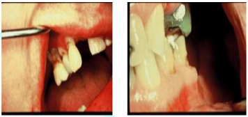
There are four distinct types of deformations that identify DCS: a non-carious wedge shaped lesion found at the gingiva (Figure 1), the inverted cupola, another Non Carious Lesion (NCL) found at the tips of functional cusps (Figure 2), exostosis (Figure 3); and deformations of restorative materials (Figure 4). Lets look at each.
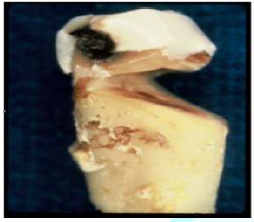
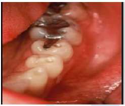
Figure 2: Inverted Cupolas–NCLS Due To Compression, Static or Cyclic Loading.
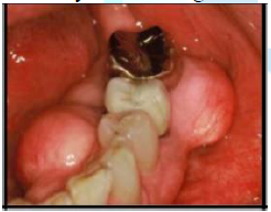

Figure 4: Luder Lines in Amalgam Due to Stress.
Gingival Ncls
A wedge shaped lesion that usually occurs at the Dentinal Enamel Junction (DEJ) of teeth. It is classified as a Non-Carious Lesion (NCL). (Figure 5) What causes it? This unique loss of tooth substance has been the subject of controversy among dentists for almost one hundred years. W.I. Ferrier once wrote; ‚their etiology seems to be shrouded in mystery [16]. But it is not such a mystery if we understand the science of biomechanics, ie; the study of the mechanical behavior of living materials and structure. What we are actually seeing are multi-shaped examples of fatigue due to compression and tension (Figure 6-13).
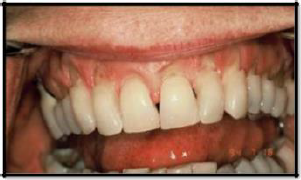
Figure 5: NCLS at the Gingiva Due to Compression and Tension.
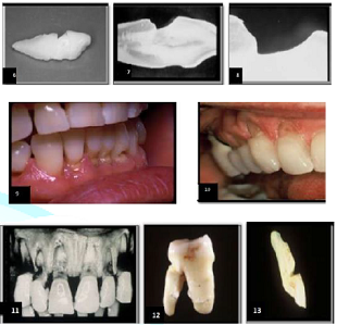
Figures 6-13: Various Examples of NCLS Due to DCS.
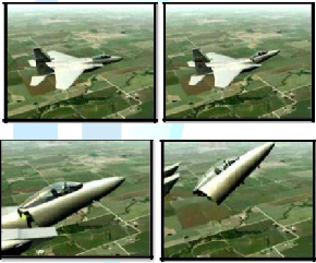
Figure 14-17: Structural Failure of Jetfighter Due to Corrosion Fatigue.
Fatigue applies to changes in the properties of a material due to repeated applications of stress or strain-in this case, compression failure from DCS. J.E. Gordon, a professor of materials at Reading University, U.K., describes fatigue as one of the most insidious causes of loss of strength in a structure [17]. If an object, such as a tennis ball, rebounds to its original shape after repeated compressions, it is said to be elastic in nature. However, if an object exhibits residual defects after repeated compression, it is said to be plastic in nature. Biological structures, such teeth and bone are termed viscoelastic and are subject to deformation. Engineers refer to this type of fatigue as corrosion fatigue.
Figures 14-17 show a jet fighter whose Cock pit was sheared from the body of the plane. The cause is corrosion fatigue which is the reduction by corrosion, of the ability of the metal to withstand cyclic or repeated stresses. So, why dont we recognize this? This is an engineering problem and the mechanisms of engineering are not emphasized in dental school because engineering is taught in high math-calculus. In dental school, we drop high math and focus more on chemistry.
Since teeth do work, it behooves us to examine the design aspects of the dentition from an engineering point of view. In 1975, a team of engineers at the University of California, Los Angeles, used the finite element method to study stress generated in a premolar as a result of occlusal forces [18]. This method is a mathematical technique which is well suited to the analysis of stress in teeth and dental restorations because it can closely simulate the geometrics, loads, and material in homogeneities in the system being studied.
The analysis (Figure 18) definitely revealed that the DEJ was susceptible to cleavage or failure planes. In this example, a failure plane is apt to occur on the lingual face running through the DEJ well down into the root-the classic geometry of a gingival notch. A year later, another team of engineers at Brunel University, Middlesex, England, did a similar study with supportive results [19]. (Figure 19)
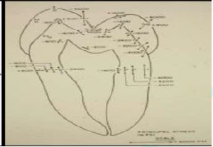
Figure 18: Magnitude and direction of principal stresses at selected locations.
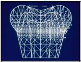
Figure 19: Axissymmetric finite element model with three marginal configurations.
Again using the finite element method to demonstrate stress distribution, these engineers concluded the tensile forces in the gingival area are powerful enough to pull apart the enamel prisms. They also concluded these high stresses are probably responsible for the pain often experienced by patients who have received cervically placed restorations. This was in the pre-bonding era where we would mechanically lock in our restorations. While these two engineering reports began to reveal valuable information about the amount of activity in the gingival zone, it was an earlier work that demonstrated the unusual flexibility of teeth. In 1968, Dr. J.A. Hood published an article in the New Zealand dental journal, experimental studies on tooth deformation [20]. Using the method of photoelasticity (Figure 20) this researcher placed teeth in a loading frame and applied pressure. The frozen stress technique utilized demonstrated an actual shortening of the tooth occlusal-gingivally with an increase in its bucco-lingual diameter.
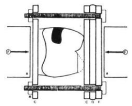
Figure 20: Compression of Tooth in a Loading Frame to Demonstrate Flexibility of teeth.
Conclusion
Teeth are flexible-a subject rarely discussed. The compression failure of an object occurs at its most vulnerable site. Teeth are most susceptible at the gingival area. If the alveolar bone recedes, the failure site will also be lowered. Figures 21 & 22 demonstrate defects that appear in tandem as the supporting bone atrophies thus changing the fulcrum point. Also note in Figure 22 that the only occlusal contact is on the incline plane, forcing the bicuspid to be flexed toward the lingual when the patient clenched. Compression fatigue can also occur in the spine (Figure 23). In orthopedics, these sites of destructive stress are termed compression or wedge fractures [21]. Sites of compression fatigue in biological structures are not uncommon and easily found in professional athletes such as ice skaters or runners, so it is unfortunate that many in the dental profession are uncomfortable with their engineering etiology.
Figures 21 and 22: Gingival Fatigue in Tandem.
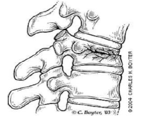
Figure 23: Vertebral Compression or Wedge Fracture.
The Inverted Upola
The most common compression NCL is a perfectly rounded depression sometimes called occlusal dimples, found at the tips of functional cusps (Figure 24). Nothing of significance has been written about them except that they are associated with compression. Although the inverted cupola contrasts dramatically in geometric design with the wedge shaped NCL, there are two striking similarities, ie; they are both site specific in that they are found at sites of high stress on teeth, and they both exhibit a glassy sheen [22]. Kornfeld wrote about this phenomenon in 1932 when he observed that these defects were hard, smooth, and glasslike in appearance [23]. The author suggests that this glassy effect is due to the exit of positive ions produced by the compression of appetite crystals in the dentition and the alveolar bone [24]. This is the result of the piezoelectric effect. A piezoelectric substance is one that produces an electric charge when a mechanical stress is applied causing the substance to be squeezed or stretched. Conversely, a mechanical deformation is produced when an electric field is applied. Electricity is composed of both negative and positive ions. It is suggested that positive ions are being emitted through these focal points of high stress carrying with them minute particles of tooth structure. This would not only explain the glassy effect, but the loss of tooth structure as well. It is not unusual to find these glassy concavities on the first molars only. The explanation is that these molars appear first in the transgression from deciduous to secondary dentition and receive the full force of bruxism that is common during this period. The compression forces on the six year molars are reduced with the emergence of the remaining dentition.
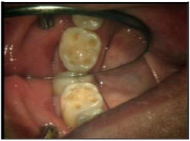
Figure 24: Compression NCLS-Tips of Functional Cusps.
Note: It is the stress which results from the various loading forces such as compression, tension, flexion, and shearing that causes tooth degradation and its effects on bone and materials. Keep in mind the equation S=F/A, where stress is concentrated, damage will occur J. Grippo, DDS.
Deformation of Bone-Exostosis
Articles on torus palatinus and torus mandibularis have appeared since 1814 [25] (Figures 25-28). Although there is no consensus as to their etiology, many associate their occurance with TMDs and masticatory hyperfunction [26,27]. Specifically, the negative ions generated from the compression of appetite crystals are responsible for the aggregates of new bone growth. This may well explain the metallic taste that people experience from time to time.
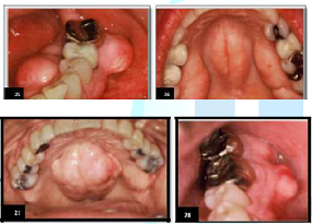
Figures 25-28: Examples of exostosis due to piezzoelectric effect.
Deformations of Restorative Materials
Fatigue easily manifests itself in prostheses and restorative materials such as amalgam and acrylic. In engineering, these wavy patterns are called‚ Luder Lines, or molecular slip bands. The explanation for the patterns is that molecules in the alloy are rearranging themselves under the influence of compressive strain. One can demonstrate the effect by bending a metal coat hanger back and forth and examining the stress configuration that is produced. Figures 29-32 demonstrate Luder Lines in restorative materials.
Epidemiology
A survey was taken of 100 patients (50 female; 50 male; age range, 17-76) to determine how many exhibited signs and symptoms of DCS and TMD [28] (Table 1). The deformations in the oral environment are important diagnostic tools, but their appearance does not mean that the patient is currently affected with DCS, as it may have been from a prior stressful period in their lives.
Management of Bruxism
The stomatognathic system best serves the patient when it functions vertically and is free from clenching and grinding. If DCS does occur, it is the obligation of the G.P. to recognize the signs, consult with the patient on management, ensure that the occlusion (the way the teeth touch each other in closure) is correct, and provide a guard if necessary. If clenching occurs during waking hours, it is the responsibility of the patient to monitor themselves and make a conscious effort to keep their teeth apart (mandible at rest). If clenching and/or grinding do occur while sleeping, it is the GPs responsibility to provide a comfortable guard. The main reason for the occlusion confusion is that we have been accommodating the horizontal component of DCS instead of trying to prevent it. We have gone from group function, to cuspid rise, and then to anterior guidance which in my opinion, doesnt make any sense at all. Anterior guidance, aka mutually protected occlusion, aka mutually protected articulation according to the Journal of Prosthetic Dentistry is defined as: An occlusal scheme in which the posterior teeth prevent excessive contact of the anterior teeth in maximum intercuspation, and the anterior teeth disengage the posterior teeth in all mandibular excursions. The general understanding of anterior guidance is that there is some sort of mutual protection at work here. Nothing could be further from the truth. Lets examine this concept in detail.

Table 1: Signs and Symptoms of DCS and TMD.
The posterior teeth prevent excessive contact of anterior teeth in maximum intercuspation
First of all, why the excessive contact? We are talking about the vertical form of DCS-clenching. If the patient is clenching vigorously during waking hours, it is their responsibility to monitor themselves and control it. If they are clenching with force while sleeping, a guard will suffice. Is it possible that clenching on the posterior teeth can prevent excessive contact on the anterior teeth? Not necessarily. A posterior tooth will be subject to more stress upon clenching than an anterior tooth simply because there is more surface contact upon closure, i.e., stress is a result of pounds per contact unit squared. However, if the occlusal contact upon closure for both molars and the lingual of the upper anteriors is equal, so will be the stress.
The anterior teeth will disengage the posterior teeth in all mandibular excursions
The idea being that if the patient is grinding, the posterior teeth will not wear. This is not logical thinking and it is harmful. Lets be clear. This is not normal function, this is parafunction. Im sure there is the exception to the rule, but generally people do not grind their teeth during waking hours, they clench, and since grinding in protrusion occurs only while sleeping, a comfortable guard would be the appropriate protection. This mutual protection theory is flawed in that it does not address the damage incurred to the anterior teeth during the parafunction. If this concept were credible, there would be little use for guards.
Consequences of the Confusion How Has the Confusion Affected the Way Dentistry is Currently Being Produced? Restorative dentistry takes place on two distinct levels; maintaining the status quo and rehabilitation/reconstruction. Lets look at each. Maintaining the Occlusal Status Quo Benchmark: The Dentition
What is the current situation?
Universally, 99% of restorative dentistry is done by increments–replacing old or creating new restorations to maintain the occlusal (The System) status quo. If the patient has no complaints regarding occlusion (How the teeth touch), the TMJ, or orofacial pain, the dentist proceeds with the restorative project. If that project, say a crown or a bridge, opposes a dentition that is naturally sharp (occlusal surface), the new restoration will correspondingly match its antagonist. A patient with a flattened dentition will likewise receive a matching restoration. If there are no interferences in the patients natural closure or lateral excursions, the new restoration is delivered. What is wrong with this scenario? What is wrong is that we have the perception that any crown design is acceptable as long as there are no interferences. This is a misconception. From an engineering point of view, increased occlusal contact (flattening) during closure will result in increased stress at the DEJ and widening of the envelope of function. In addition, the subsequent loss of the intra incline space will limit anterior-posterior movement of the mandible during postual changes.
Is there a better way?
We have two objectives when restoring a segment of a patients dentition. The first is to correctly design the new restoration from an engineering point of view, and the second is to ensure that it is in harmony with healthy function of the stomatognathic system.
How is this done?
We have a unique problem on our hands; part engineering and part psychological. This means we have to accomplish three things;
1. Design the new restoration to satisfy good engineering principles with the occlusal contact at the tip of the functional cusp and touchless incline planes.
2. To evaluate the stomotognathic system itself for signs of DCS. If the patient is affected with parafunction the dentist must work with the patient on management.
3. To evaluate the remaining dentition to determine whether an equilibration might be indicated.
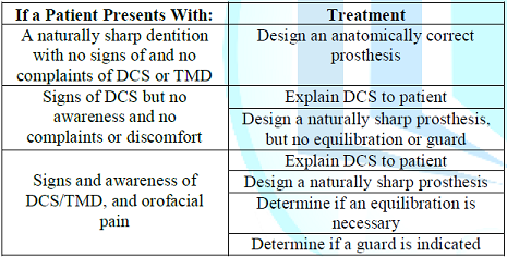
Table 2: Patient complaints and suggested treatment.
Here are some general rules
Management of DCS begins with the recognition of the deformations of the dentition, bone, and restorative materials in the oral environment. This should initiate a dialogue between the dentist and the patient as to the awareness of the problem since bruxism works well within the subconscious. So a light discussion, not an accusation is in order. It should be noted that the observed deformations may have occurred during a prior time period of stress and the patient may be currently comfortable. If the patients occlusion (occlusal contact during closure) is uncomfortable, it should be determined whether an equilibration is indicated.
The equilibration
At the American Equilibration Societys (AES) 56th meeting in Chicago, there were four speakers; John Kois, Clayton Chan, Christopher Orr, and Glenn DuPont, each demonstrating four different methods of equilibration. They all had good results. Which is the best and simplest method of equilibration for a GP to use when doing piece meal restorative dentistry? I dont believe it is necessary to use complicated instrumentation to accomplish an equilibration. I simply apply occlusal indicator wax on the occlusal surfaces of one arch and have the patient squeeze once. The areas of displaced wax are analyzed. Where are they? Are they at the tip of the cusp or on the incline planes? Are they large or small? Ideal contacts are small and confined to the tips of the cusps and the central fossae. If the incline planes are touching, I reduce them. If there is heavy contact in the central fossae I sharpen the opposing functional cusps. I do check their lateral excursions for interferences, but I warn patients not to go into these harmful lateral compressions. The teeth are never shortened. The feeling after an equilibration is one of ‚less. The equilibration reduces the stress on the teeth and allows greater freedom for the mandible, however it doesnt mean the patient wont clench if stressed-thats where the guard comes in.
The exception to the rule: the original morphology of our teeth is superior to that of a flattened dentition. However, I dont mean to imply that all flat teeth should be equilibrated (sharpened). I have seen many patients over the years with zero degree occlusion, and in most cases they are quite comfortable. The damage has been done. To equilibrate their teeth would be disastrous. However there should be a general understanding that preserving the original morphology of the dentition is superior and more efficient than a flattened dentition. If patients are actively compressing their teeth while sleeping, then a guard is mandatory. It is normal for many patients to grind and clench while sleeping. It just means that their minds are very active or they are dreaming and the purpose of the guard is not to get them to stop, but to provide an acrylic spacer to absorb the compressive forces. I once witnessed two poster board displays at an AES meeting prepared by the graduate students at Northwestern University Dental School. One was entitled ‚Guards Work‛ and listed all the benefits of guard therapy. The other poster board was entitled‚ Guards dont work and had a list of problems that could occur by wearing them. So, whats the point? The point is that you have to find the right guard for the right patient. I personally endorse the smaller anterior hard guards as they reduce the compression force by 75%, but others recommend the larger guard covering the molars. They point out that their purpose is to support the TMJ. To my way of thinking it only adds more stress to the TMJ. So here is another contributing factor to the confusion that needs debate.
Note: Soft guards although comfortable, may encourage clenching and guards should only be worn while sleeping. Excessive use of a guard during waking hours can cause micro movement of the teeth.
Rehabilitation/Reconstruction
A small percentage of dentists limit their practices to patients who are severely compromised with problems relating to occlusion (Stomatognathic System). There are many post graduate study groups that are dedicated to understanding and solving these problems, the most noted being the Las Vegas Institute (LVI), The Dawson Group, and the Pankey Institute. While there is some professional competition between these groups as to which methodology is superior, they all are very successful, depending of course, on the skill and experience of the dentist. The percentage of dentists dedicated to rehabilitation is estimated at less than 1%.
The Neuromuscular Concept
Benchmark:
Muscles of Mastication
Using Transcutaneous Electric
Nerve Stimulation (TENS), the goal of neuromuscular rehabilitation is to first
establish a physiologic terminal contact position, i.e.; the myocentric bite.
The incline planes of the teeth are then refined to ensure physiologic
mandibular function. Stabilization of the masticatory system is
achieved by using a removable anatomical orthotic appliance in which cuspid
rise is incorporated.
The Gnathological Approach
Benchmark:
Position of the Condyles in the Glenoid Fossae
The goal of the gnathologist in
the reconstruction/rehabilitation of the stomatognathic system is to obtain an
optimum orthopedically stable joint position. It is called Centric Relation (CR),
and is defined as the position of the condyles when they are in the
anterior-superior position in the glenoid fossae resting against the posterior
slopes of the articular eminences, with the articular discs properly
interposed. This position is considered to be the most musculoskeletally
stable position of the mandible. It is also the objective of the gnathologist
to have stable holding contacts on all teeth to support the condyles in this
centric relation. This position is termed Centric Occlusion (CO). The ultimate
objective gnathological
approach is to establish long-term occlusal stability. This means, as
Okeson explains, to establish an occlusal condition that can accept heavy
forces with minimal damage and at the same time be functionally efficient.
Discussion
While this all sounds credible and reasonable, the neutral observer has to ask about the fortification of the stomatognathic system in order to accept heavy forces. What heavy forces? Arent we talking about the vertical and horizontal forces of DCS? And since these heavy forces only occur while sleeping-why not just make them a guard? Again, we are accommodating DCS rather than trying to prevent it.
In my opinion
We have made this subject far more complicated than it has to be. We have so many conflicting ideas and theories and practical concepts that a majority of GPs are unsure about what the right approach should be toward restoration. One unfortunate legacy of the confusion is that we really dont agree as to the best design of teeth. It is important that teeth retain their original morphology in order to work harmoniously so as to not distract the mandible from normal function. Regrettably, this third, equally important benchmark has been largely ignored. In fact, the role of the dentition in the function and dysfunction of the system has been obfuscated to the point that many believe there is no relationship at all. Ramfjord and Ash reported in 1995; ‚a trend of thinking has developed that virtually denies any relationship between occlusal (the way teeth touch in closure) factors and disorders of the masticatory system [29]. Unfortunately, this attitude is prevalent today. The question is how it could not?
Preserving the human dentition in its natural state is critical to maintaining the health and efficiency of the system. The prevention of fatigue failure by good design is critical. Some think design means how it looks, but its really how it works. Maintaining the original design of the dentition will enable you to produce superior dentistry. We must examine the morphology of teeth with a fresh point of view. Vertical function with a naturally sharp dentition is healthy and efficient. The idea that a flattened occlusal surface is acceptable is outdated dogma. Sharp teeth are superior. It is time to look beyond the horizon of antiquated views.
What can we do to clarify the confusion?
An important step would be to stop using the word occlusion in such a broad sense. Being more specific will simplifies the thought or question. For instance, lets look at two questions that have plagued us for the past 80 years.
What is an ideal occlusion?
Turp et al. discuss the quest for the ideal occlusion [30]. They explain that an ideal occlusion is rarely found in real life, that the idea is open to personal interpretation, and that it is presumptuous to state natures intention for idealism. I totally disagree. We dont need a clairvoyant to tell us what ideal is, we just need a bioengineer with some knowledge of physiology. We also need to be specific about what is meant by the question. If we are concerned about the stomatognathic system, an ideal system, no matter class I, II, or III, would be one that is free from parafunction. The teeth in this parafunctionally free system, no matter crooked or straight, are naturally sharp, fit loosely with their antagonists, and have the occlusal contact confined to the tip of the cusp upon closure.
What is the relationship between occlusion and TMJD?
Again, the different interpretations of the word occlusion make this question difficult to answer. Lets rephrase the question to: what is the relationship between the stomatognathic system and TMJD? It is recognized that problems of the TMJ can be the result of trauma, developmental deformities, or a disease process, but in my opinion, the vast majority of TMJ problems are the result of repetitive motion trauma from DCS. In my 40+ years of clinical practice, I have yet to witness a single case of TMD that did not have at least one or more signs of parafunction. So a better question would be; what does parafunction have to do with problems with the TMJ and then determine whether the occlusal contacts are a contributing factor.
A call for consensus
Every five years, the Japanese sponsor an international consensus meeting on implantology. Respected authorities are invited to express their views on important issues and a panel of judges determines a consensus for each issue for that point in time. Isnt it about time we had one on occlusion? Until there is a consensus, it is suggested that general practitioners follow a simple theme of keeping teeth sharp and keeping them apart.
References
1. Christensen GJ. Personal Correspondence. 11 06, 2008.
2. Dorland W. Dorlands Pocket Medical Dictionary 1st Edn (1898) WB Saunders, Philadelphia.
3. Dorlands Pocket Medical Dictionary 20th Edn (1959) WB Saunders, Philadelphia
4. Jankelson B, Hoffman GM and Hendron, JA. Physiology of the Stomatognathic System (1953) J Am Dent Assoc 1953 46: 375. https://doi.org/10.14219/jada.archive.1953.0070
5. Rugh JD and Smith BR. A testbook of occlusion (1988) Quintessence Publishing Co, Chicago 143.
6. Jablonski S. Illustrated dictionary of dentistry (1982) WB Saunders, Philadelphia.
7. Okeson JP. Management of temporomandibular disorders and occlusion (1993) CV Mosby, USA 51.
8. Okeson JP. Management of temporomandibular disorders and occlusion 3rd Edn (1993) CV Mosby, USA 125.
9. McCoy G. The truth about occlusion (2007) Presented at Yankee Dental Congress San Francisco.
10. Ortman HR. Essentials of complete denture prosthodontics, 2nd Edn Winkler IN (ed) (1988) PSG Pub. Co, Mosby, USA, 464.
11. Kydd WL. Complete Denture Base Deformation with Varied Occlusal Tooth Form (1956) J Prosthet Dent 6: 714-718. https://doi.org/10.1016/0022-3913(56)90018-X
12. Regli CP and Kydd WL. A preliminary study of the lateral deformation of metal base dentures in relation to plastic base dentures (1953) J Prosthet Dent 3: 326-330. https://doi.org/10.1016/0022-3913(53)90007-9
13. Swoope CC and Kydd WL. The effect of cusp form and occlusal surface area on denture base deformations (1966) J Prosthet Dent 16: 34-43. https://doi.org/10.1016/0022-3913(66)90110-7
14. Neilburger E. Flat plane occlusion in the developement of man (1977) J Prosthet Dent 38: 459-469. https://doi.org/10.1016/0022-3913(77)90101-9
15. McCoy G. On the Longevity of Teeth (1983) J Oral Implan 2: 248-267.
16. Ferrier WI. Clinical observations on erosions and their restorations (1931) J California State Dent Assoc 20: 1150-1163. https://doi.org/10.14219/jada.archive.1933.0206
17. Gordon JE. Structures or Why Things Dont Fall Down (1978) Da Capo Press, USA 333-334.
18. Selna LG, Shillingburg HT and Kerr PA. Finite element analysis of dental structures-axisymmetric and plane stress idealization (1975) J Biomed Matter 9: 237-252. https://doi.org/10.1002/jbm.820090212
19.
Yettram
AL, Wright KWJ and Pickard HM. Finite element stress analysis of the crowns of
normal and restored teeth (1976) J Dent Re 55.
https://doi.org/10.1177/00220345760550060201
20. Hood, JAA. Experimental Studies on Tooth Deformation in Class Five Restorations (1972) New Zealand Dent J 116-131.
21. Old JL and Calvert M. Vertebral compression fractures in the elderly (2004) Am Family Physician 69: 111-116.
22. McCoy G. Examining the Role of Occlusion in the Function and Dysfunction of the Human Masticatory System (1997) Nippon Dent Rev 659: 163-183.
23. Kornfeld B. Preliminary report of clinical observation of cervical erosions (1932) Dent Items Interest 54: 905-909.
24. McCoy, G. Dental Compression Syndrome and TMD, Examining the Relationship (2007) Dent Today 26: 118-123.
*Corresponding author
Gene McCoy, San Francisco, California, 94123, USA, E-mail: genemccoydds@sbcglobal.net
Citation
McCoy G. Occlusion confusion (2019) Dental Res Manag 3: 16-23
